. 2004 Mar;78(5):2405-13.
doi: 10.1128/jvi.78.5.2405-2413.2004.

The DNA-A component of a plant geminivirus (Indian mung bean yellow mosaic virus) replicates in budding yeast cells

Affiliations

The DNA-A component of a plant geminivirus (Indian mung bean yellow mosaic virus) replicates in budding yeast cells

Vineetha Raghavan et al. J Virol. 2004 Mar.

Abstract

Understanding the biochemistry of DNA replication of the plant DNA viruses is important for the development of antiviral strategies. Since DNA replication is little studied in plants, a genetically tractable, easily culturable, eukaryotic model system is required to pursue such studies in a facile manner. Here we report the development of a yeast model system that supports DNA replication of a chosen geminivirus strain, Indian mung bean yellow mosaic virus. The replication of plasmid DNA in the model system relies specifically on the virus-derived elements and factors. Usage of this model system revealed the role of at least one hitherto unknown viral factor for viral DNA replication. The episomal characteristic of single-strandedness of replicated plasmid DNA was shown, and the expression of viral genes was also confirmed. This model system is expected to shed light on the machinery and mechanism involved in geminiviral DNA replication in plants.


Figures
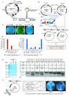
FIG.1.
FIG.1.
Genetic studies on geminiviral replication in yeast. (A) Genome organization of IMYMV Black-gram isolate (IMYMV-Bg) DNA-A. An enlargement of the characteristic hairpin loop region containing the conserved nonamer sequence TAA TAT TAC where Rep initiates RCR (Rep cutting site [arrow]) is shown to the right. The various ORFs with their directions of transcription are shown. The putative Rep-binding sites are underlined. The unique sites of BamHI and HindIII are marked. (B) Strategy used to make the replication-competent virus recombinant plasmid YCpO-2A. The nomenclature YCpO-2A-For or -Rev was on the basis of orientation of the AV1 ORF with respect to the AmpR gene in YCpO. (C) Representative plates comparing colonies obtained of the transformed yeast W303a strain (MATa leu2 ura3 his trp1 ade2) with plasmid YCp50 (i), YCpO (ii), or YCpO-2A (iii). (D) Panel i, % origin activity of the various constructs relative to YCp50, which was given a value of 100; panel ii, mutation of different ORFs in the DNA-A (in the YCpO-2A background), namely, AC1, AC3, AC4, and AC5, led to a decrease in origin function (% relative origin activity) compared to plasmid YCpO-2A, which was given a value of 100 in this case. The superscripts T and TF stand for termination and termination followed by frameshift mutations, respectively. (E) Panel i, SDS-polyacrylamide gel electrophoresis profile showing the apparent homogeneity in preparations of recombinant Rep and its mutant proteins. AC4T and L62I-Rep represent the same mutant. Panel ii, in vitro site-specific cutting activity of various recombinant versions of Rep with either a wild-type (Ori Wt) or mutant (OriM) oligonucleotide (sequences are indicated at the top). The 43-kDa Rep Wt was able to efficiently cut the 5′-labeled (*) Ori-Wt oligonucleotide (lane 2), and this site-specific nicking was competed out by 40× unlabeled Ori Wt (lane 3), but not by OriM (lane 4). 1× oligonucleotide represents about 1 ng of the 26-mer oligonucleotide. (F) cdc6 complementation. cdc6-YEp352 and YCpO-2A plasmids were used as a positive and negative control, respectively. The plasmids used for transformation are shown in the sector diagram. All colonies were scored on Ura dropout plates.
FIG. 2.
FIG. 2.
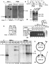
Southern blot and PCR analyses of plasmid products replicated in the W303a yeast strain. (A) Southern blotting of replicated plasmids. Plasmid DNA was isolated from transformed yeast by using a standard protocol (11) or from E. coli DH5α and was treated with a unique cutter, such as HindIII, XhoI, or BglII (H-3, X1, or B2). The digested DNA was resolved by electrophoresis in a 0.7% agarose gel, Southern blotted, and probed as shown. A 2.7-kb IMYMV DNA-A fragment obtained by HindIII digestion of plasmid pUC18-A was used as a length standard (lane 1). (B) Comparison of S1 nuclease sensitivities of plasmid DNAs derived from bacterial and yeast sources. S1 nuclease was used at a concentration of 2 U/μg of DNA at 37°C for 30 min. (C) Characterization of single-strandedness of plasmids replicated in yeast. For further confirmation of the presence of single-stranded DNA, 5 μg of YCpO-2A plasmid DNA isolated from either E. coli DH5α or S. cerevisiae W303a was allowed to bind separately to mini-HAP columns and step eluted with phosphate buffer (pH 6.8) as indicated. M13 single-stranded DNA and double-stranded RF markers were used as standards during HAP chromatography and were eluted with 75 to 150 mM and 220 to 350 mM phosphates, respectively. The different fractions of eluted plasmids were either mock treated or treated with S1 nuclease. The DNAs of various fractions were sufficiently resolved by agarose gel electrophoresis, Southern blotted, and autoradiographed. The slower migrating bands represent multimeric forms of the 12.5-kb YCpO-2A. The quantitation of DNA was carried out by measuring the intensities of the 12.5-kb DNA bands in the appropriate lanes with Kodak ID2.0 software. The single-stranded DNA-containing fractions were derived from lanes 11 and 13 (without S1) and 12 and 14 (with S1). Similarly, only the double-stranded DNA fractions from the yeast source were recovered from lanes 15 and 17 (without S1) and 16 and 18 (with S1). The positions of the 12.5-kb DNAs are marked by arrows. (D) Integrity of double copies of DNA-A during replication in yeast. Panel i, the DNA present at the HindIII site of the yeast-derived plasmids was amplified by using primers YCpFwd and YCpRev. The purified 5.6-kb amplified product (lane 1) was digested with BamHI (position 960) to obtain three fragments, of 2.7 kb (a), 1.8 kb (b), and 1.1 kb, respectively (lane 3). The top diagram shows the locations of CRs in the BamHI fragments. Panel ii, fragments a and b containing one origin (CR) were each purified and used as templates for amplification with duplex PCR primer combinations as indicated in the table. For each amplification, two bands of the expected sizes were obtained. The relative positions of the primers employed are shown at the top. The IMYMV DNA-A was used as control template C. (E) Model indicating possible modes of replication of YCpO-2A plasmid in yeast. We hypothesize that only one of the origins, either ori1 (i) or ori2 (ii), is able to initiate RCR generating full-length YCpO-2A.
FIG. 3.
FIG. 3.
Viral transcripts in S. cerevisiae bearing plasmid YCpO-2A and in infected French bean plants. (A) RT-PCR from RNAs of yeast samples transformed with various plasmids. The PCR products of cDNAs derived from yeast samples (4) bearing YCpO-2A plasmids are shown for the pairs of primers specific for the Rep (lanes 10 and 11), AC3 (lane 12), and CP (lane 13) genes of IMYMV. RepC is the C-terminal part of Rep. The expected pattern of amplification is shown in lanes 2 to 5 of the positive (+ve) control PCR panel that was obtained from direct amplification from a template of viral DNA-A. The cDNA prepared from the total RNA extracted from YCp50 plasmid-bearing yeast was used as a negative control (lanes 6 to 9). A negative (−RT) control is shown in lanes 14 and 15 indicating that the PCR products are not the artifacts of DNA contamination in the processed RNA samples. A nonspecific band at around 550 bp was observed for both YCp50 control and YCpO-2A samples in the case of AC3 primers (lanes 8 and 12). Southern blotting with an actin probe was carried out to show uniformity in the loading of cDNA templates (bottom panel). (B) Northern blot analysis of total RNA extracted from the yeast strain W303a bearing the YCpO-2A plasmid. The presence of a 1.8-kb Rep transcript was revealed by probing with radiolabeled AC1 DNA. Such a transcript was absent from the RNA samples of yeast transformed with the YCp50 control plasmid, as expected. This result is consistent with the observation made with IMYMV-infected French bean plants (C). The total RNAs isolated from the appropriate sources are shown at the bottom for panels B and C.
FIG. 4.
FIG. 4.
Expression of the viral CP in S. cerevisiae transformed with YCpO-2A. (A) Western blot analysis of IMYMV CP. The total protein was prepared from untransformed and transformed yeast by resuspension of the cell pellet from 1 ml of culture, obtained at the mid-log phase, in 1× sample buffer (0.06 M Tris-HCl [pH 6.8] 10% [vol/vol] glycerol, 2% [wt/vol] SDS, 5% [vol/vol] 2-mercaptoethanol, 0.0025% [wt/vol] bromophenol blue) and boiling for 10 min. The plasmids used for transformation are indicated at the top of each lane (lanes 2 to 4). Fifty micrograms of each protein sample was separated in an SDS-10% polyacrylamide gel and immunoblotted with a heterologous Indian cassava mosaic virus CP antibody. Three hundred nanograms of purified maltose binding protein fusion of CP (MBP-CP) was used as a positive control. (B) Confocal imaging of yeast cells bearing the YCpO-2A plasmid. The CP was expressed throughout the yeast cells (b and e), and the recognition specificity of the CP antibody was highlighted by the absence of CP staining in the control cells harboring plasmid Ycp50 (h). Panels a, d, and g show nuclei stained with DAPI, and panels c, f, and i show transmission images in white light. Bars, 1 μm. The same sets of cells are shown in rows, and cells at different growth stages are shown in columns.

Similar articles

Cited by

References

    1. Ach, R. A., T. Durfee, A. B. Miller, P. Taranto, L. Hanley-Bowdoin, P. C. Zamryski, and W. Gruissem. 1997. RRB1 and RRB2 encode maize retinoblastoma-related proteins that interact with a plant D-type cyclin and geminivirus replication protein. Mol. Cell. Biol. 17:5077-5086. - PMC - PubMed
    1. Agatep, R., R. D. Kirkpatrick, D. L. Parchaliuk, R. A. Woods, and R. D. Gietz. 1998. Transformation of Saccharomyces cerevisiae by lithium acetate/single stranded DNA/polyethylene glycol (LiAc/ss-DNA/PEG) protocol. Technical Tips Online [Online.] http://tto.trends.com.
    1. Angeletti, P. C., K. Kim, F. J. Fernandes, and P. F. Lambert. 2002. Stable replication of papillomavirus genomes in Saccharomyces cerevisiae. J. Virol. 76:3350-3358. - PMC - PubMed
    1. Ausubel, F. M., R. Brent, R. E. Kingston, D. D. Moore, J. G. Seidman, J. A. Smith, and K. Struhl. 1998. Current protocols in molecular biology, vol. 2. John Wiley & Sons, Inc., London, United Kingdom.
    1. Castillo, A. G., D. Collinet, S. Deret, A. Kashoggi, and E. R. Bejarrano. 2003. Dual interaction of plant PCNA with geminivirus replication accessory protein (Ren) and viral replication protein (Rep). Virology 312:381-394. - PubMed

Publication types

Substances

LinkOut - more resources