. 2004 Mar;78(5):2517-29.
doi: 10.1128/jvi.78.5.2517-2529.2004.

Inhibition of human immunodeficiency virus type 1 replication by RNA interference directed against human transcription elongation factor P-TEFb (CDK9/CyclinT1)

Affiliations

Inhibition of human immunodeficiency virus type 1 replication by RNA interference directed against human transcription elongation factor P-TEFb (CDK9/CyclinT1)

Ya-Lin Chiu et al. J Virol. 2004 Mar.

Abstract

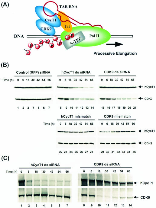
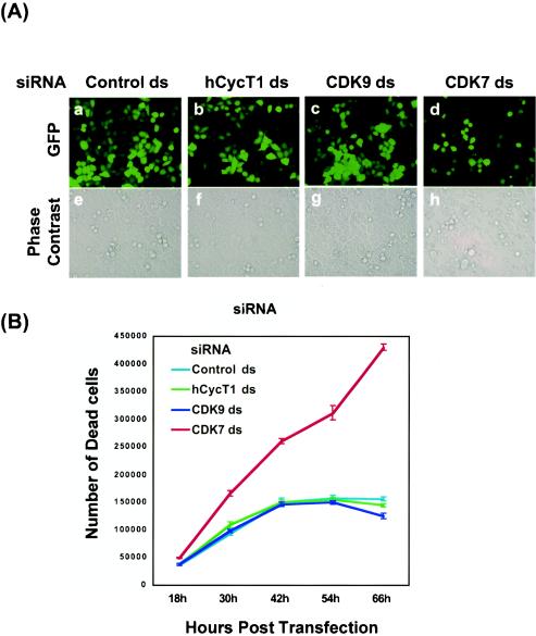
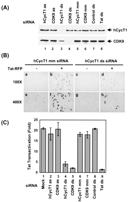
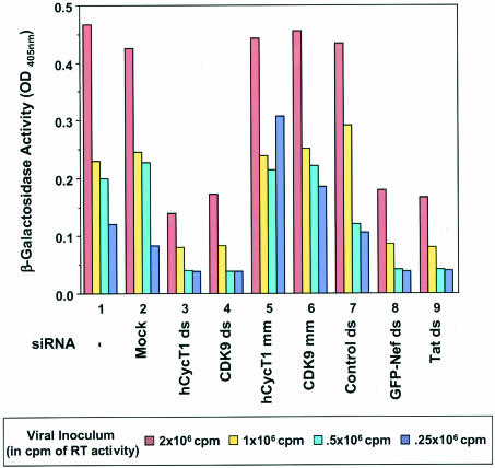
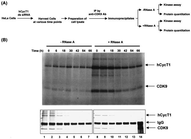
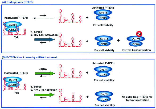
The human positive transcription elongation factor P-TEFb is composed of two subunits, cyclin T1 (hCycT1) and CDK9, and is involved in transcriptional regulation of cellular genes as well as human immunodeficiency virus type 1 (HIV-1) mRNA. Replication of HIV-1 requires the Tat protein, which activates elongation of RNA polymerase II at the HIV-1 promoter by interacting with hCycT1. To understand the cellular functions of P-TEFb and to test whether suppression of host proteins such as P-TEFb can modulate HIV infectivity without causing cellular toxicity or lethality, we used RNA interference (RNAi) to specifically knock down P-TEFb expression by degrading hCycT1 or CDK9 mRNA. RNAi-mediated gene silencing of P-TEFb in HeLa cells was not lethal and inhibited Tat transactivation and HIV-1 replication in host cells. We also found that CDK9 protein stability depended on hCycT1 protein levels, suggesting that the formation of P-TEFb CDK-cyclin complexes is required for CDK9 stability. Strikingly, P-TEFb knockdown cells showed normal P-TEFb kinase activity. Our studies suggest the existence of a dynamic equilibrium between active and inactive pools of P-TEFb in the cell and indicate that this equilibrium shifts towards the active kinase form to sustain cell viability when P-TEFb protein levels are reduced. The finding that a P-TEFb knockdown was not lethal and still showed normal P-TEFb kinase activity suggested that there is a critical threshold concentration of activated P-TEFb required for cell viability and HIV replication. These results provide new insights into the regulation of P-TEFb function and suggest the possibility that similar mechanisms for monitoring protein levels to modulate the activity of proteins may exist for the regulation of a variety of other enzymatic pathways.


Figures

FIG. 1.
FIG. 1.
Specific silencing of P-TEFb expression by RNAi. (A) Model for HIV Tat transactivation involving the human P-TEFb (CycT1-CDK9) complex. See the text for details. (B) Analysis of specific hCycT1 and CDK9 RNAi activities by Western blotting. HeLa cells were transfected with double-stranded (ds) siRNAs targeting RFP (control; lanes 1 to 7), hCycT1 (lanes 8 to 14), or CDK9 (lanes 15 to 21). Cells were also transfected with mutant siRNAs (hCycT1 mismatch [lanes 22 to 28] or CDK9 mismatch [lanes 29 to 35]) with 2-nt mismatches between the target mRNA and the antisense strand of siRNA at the hypothetical cleavage site of the mRNA. Cells were harvested at various times posttransfection. Protein contents were resolved by SDS-10% PAGE, transferred onto PVDF membranes, and immunoblotted with antibodies against hCycT1 and CDK9. (C) Analysis of specific hCycT1 and CDK9 RNAi activities by RT-PCR. HeLa cells were transfected with hCycT1 double-stranded siRNA (lanes 1 to 7) and CDK9 double-stranded siRNA (lanes 8 to 14) and were harvested at various times after transfection, and mRNA was extracted. One-step RT-PCRs were performed, with specific primers for hCycT1 and CDK9 amplification (see Materials and Methods for details). RT-PCR products were resolved in 1% agarose gels and viewed by ethidium bromide staining.
FIG. 2.
FIG. 2.
P-TEFb silencing is not lethal to HeLa cells. (A) Analysis of cell viability by in vivo fluorescence analysis. HeLa cells were cotransfected by use of Lipofectamine with a pEGFP-C1 reporter (GFP) plasmid and siRNAs (see Materials and Methods). Four siRNA duplexes, including a control duplex targeting RFP (panels a and e) and three duplexes targeting hCycT1 (panels b and f), CDK9 (panels c and g), and CDK7 (panels d and h), were used in these experiments. Reporter gene expression was monitored at 50 h posttransfection by fluorescence imaging of living cells (upper panels). Cellular shapes and densities were recorded by phase-contrast microscopy (lower panels). (B) Analysis of cell viability by counting of trypan blue-stained cells. HeLa cells were cotransfected by use of Lipofectamine with a pEGFP-C1 reporter (GFP) plasmid and siRNAs (see Materials and Methods). Four siRNA duplexes, including a control unrelated duplex (light blue) and three duplexes targeting hCycT1 (green), CDK9 (dark blue), and CDK7 (red), were used in these experiments. At various times after transfection, cells floating in the medium were collected and counted in the presence of 0.2% trypan blue. Cells that took up dye (stained blue) were not viable.
FIG. 3.
FIG. 3.
hCycT1 and CDK9 duplex siRNAs inhibit HIV-1 Tat transactivation in Magi cells. (A) Analysis of hCycT1 and CDK9 RNAi activities in Magi cells by Western blotting. Magi cells were cotransfected with pTat-RFP plasmid and various siRNAs. Cells were harvested at 48 h posttransfection. Proteins were resolved by SDS-10% PAGE, transferred onto PVDF membranes, and immunoblotted with antibodies against hCycT1 (upper panel) and CDK9 (lower panel). Lanes 1 and 2, RNAi activities in Magi cells treated with antisense (as) strands of hCycT1 and CDK9 siRNAs; lanes 3 and 4, RNAi activities of cells treated with double-stranded siRNAs targeting hCycT1 and CDK9; lanes 5 and 6, RNAi activities in cells treated with mutant hCycT1 siRNA (hCycT1 mm) or mutant CDK9 siRNA (CDK9 mm). A double-stranded GFP siRNA was used as an unrelated control (lane 7), while double-stranded Tat siRNA was used to target mRNA encoding Tat (lane 8). (B) Photomicrographs of β-Gal-stained Magi cells. Magi cells were untransfected (panels a, c, e, and g) or transfected (panels b, d, f, and h) with pTat-RFP in the presence of mismatched hCycT1 siRNA (mm) (panels b and f) or hCycT1 double-stranded siRNA (panels d and h). LTR activation (represented by β-Gal-stained cells) was reduced in the hCycT1 double-stranded siRNA-treated cells (panels d and h). (C) Effect of P-TEFb silencing by RNAi on Tat transactivation in Magi cells. Twenty-four hours after pretreatment of Magi cells with siRNA, the cells were cotransfected with pTat-RFP plasmid and various siRNAs. Cells were harvested at 48 h post-pTat-RFP transfection, and the activity of β-Gal in clear cell lysates was measured (see Materials and Methods). Tat transactivation was determined by the ratio of β-Gal activity in pTat-RFP-transfected cells to that in cells without pTat-RFP treatment. Inhibitory activity was determined by normalizing the Tat transactivation activity to the amount of Tat-RFP protein (see Materials and Methods) in the presence or absence of siRNA treatment. Bar 1, Tat-RFP transfection (mock). Magi cells were cotransfected with double-stranded siRNAs targeting hCycT1 and CDK9 (bars 4 and 5), with antisense (as) RNA strands (bars 2 and 3), or with mutant (mm) siRNAs (bars 6 and 7). Double-stranded GFP siRNA was used as an unrelated control (bar 8), while a double-stranded Tat siRNA targeting the mRNA encoding the Tat sequence was used as a positive control (bar 9). Means ± standard deviations (SD) of two experiments are shown.
FIG. 4.
FIG. 4.
siRNAs targeting CycT1 or CDK9 modulate HIV-1 replication. HeLa CD4-LTR-β-Gal (Magi) cells were transfected with homologous (ds; bars 3 and 4) and mismatched (mm; bars 5 and 6) siRNAs directed against CycT1 or CDK9. Cells were also mock transfected without siRNA (bar 2) or transfected with an unrelated double-stranded siRNA against the RFP sequence (bar 7). Sixteen hours later, cells were infected with HIVNL-GFP, an infectious molecular clone of HIV-1. Data for cells infected with virus and not treated with Oligofectamine are shown with bar 1. HIV-1 Tat-mediated transactivation of the LTR led to β-Gal production, which was quantified at 36 h postinfection. Cells treated with double-stranded siRNAs targeting Nef (bar 8; note that in this clone, Nef is fused to GFP, as previously reported [22]) and targeting the mRNA encoding the Tat sequence (lane 9) served as positive controls. Serial double dilutions of the viral inoculum (RT activity in counts per minute) are consistent with eightfold decreases in viral replication.
FIG. 5.
FIG. 5.
Evaluation of P-TEFb kinase activity in P-TEFb knockdown cells. (A) Experimental procedure for assaying P-TEFb kinase activity in cells with or without hCycT1 siRNA treatment. See Materials and Methods for details. IP, immunoprecipitation. (B) Kinase activity of P-TEFb. P-TEFb and its associated factors were affinity purified (anti-CDK9 immunoprecipitation) from HeLa cell extracts and were treated (lanes 8 to 14) or not treated (lanes 1 to 7) with RNase A as outlined in panel A. Kinase assays were performed with anti-CDK9 immunoprecipitates at 37°C for 1 h in a solution of 20 mM Tris-HCl (pH 8.0), 5 mM MgCl2, 60 mM NaCl, and 10 μM ATP and [γ-32P]ATP in a total volume of 45 μl. The reaction was terminated by the addition of 15 μl of 4× Laemmli sample buffer. Phosphorylated proteins were visualized by autoradiography after electrophoresis in an SDS-10% polyacrylamide gel (upper panel). hCycT1 and CDK9 proteins in the immunoprecipitates were eluted with SDS, resolved by SDS-10% PAGE, and stained with a Bio-Rad silver stain plus kit (bottom panel). The specificity of the protein bands was confirmed by immunoblotting with anti-hCycT1 or anti-CDK9 antibody (data not shown).
FIG. 6.
FIG. 6.
Model for how siRNA-mediated P-TEFb silencing modulates HIV-1 transcription without causing a major lethal effect on host cells. See the text for details. (A) Endogenous P-TEFb. (B) P-TEFb knockdown by siRNA treatment.

Similar articles

Cited by

References

    1. Bai, J., J. Sui, R. Y. Zhu, A. S. Tallarico, F. Gennari, D. Zhang, and W. A. Marasco. 2003. Inhibition of Tat-mediated transactivation and HIV-1 replication by human anti-hCyclinT1 intrabodies. J. Biol. Chem. 278:1433-1442. - PubMed
    1. Bieniasz, P. D., T. A. Grdina, H. P. Bogerd, and B. R. Cullen. 1998. Recruitment of a protein complex containing Tat and cyclin T1 to TAR governs the species specificity of HIV-1 Tat. EMBO J. 17:7056-7065. - PMC - PubMed
    1. Bieniasz, P. D., T. A. Grdina, H. P. Bogerd, and B. R. Cullen. 1999. Recruitment of cyclin T1/P-TEFb to an HIV type 1 long terminal repeat promoter proximal RNA target is both necessary and sufficient for full activation of transcription. Proc. Natl. Acad. Sci. USA 96:7791-7796. - PMC - PubMed
    1. Chao, S. H., K. Fujinaga, J. E. Marion, R. Taube, E. A. Sausville, A. M. Senderowicz, B. M. Peterlin, and D. H. Price. 2000. Flavopiridol inhibits P-TEFb and blocks HIV-1 replication. J. Biol. Chem. 275:28345-28348. - PubMed
    1. Chiu, Y. L., and T. M. Rana. 2002. RNAi in human cells: basic structural and functional features of small interfering RNA. Mol. Cell 10:549-561. - PubMed

Publication types

MeSH terms

LinkOut - more resources