. 2004 Mar;78(5):2545-52.
doi: 10.1128/jvi.78.5.2545-2552.2004.

Assembly properties of the human immunodeficiency virus type 1 CA protein

Affiliations

Assembly properties of the human immunodeficiency virus type 1 CA protein

Barbie K Ganser-Pornillos et al. J Virol. 2004 Mar.

Abstract

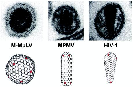
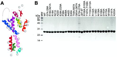
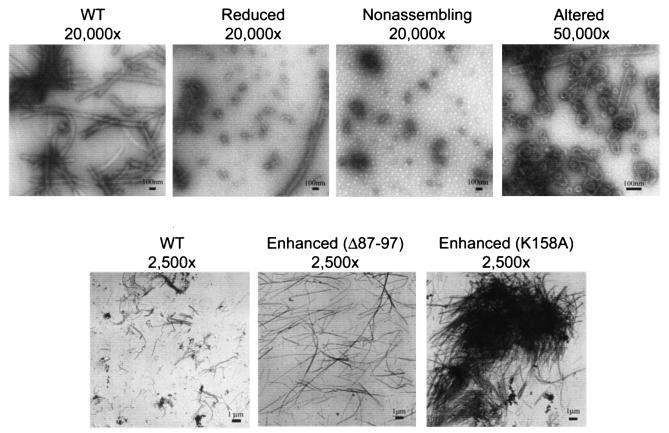
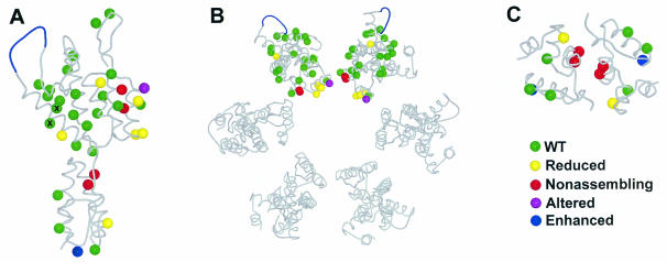
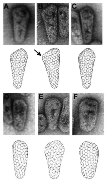
During retroviral maturation, the CA protein oligomerizes to form a closed capsid that surrounds the viral genome. We have previously identified a series of deleterious surface mutations within human immunodeficiency virus type 1 (HIV-1) CA that alter infectivity, replication, and assembly in vivo. For this study, 27 recombinant CA proteins harboring 34 different mutations were tested for the ability to assemble into helical cylinders in vitro. These cylinders are composed of CA hexamers and are structural models for the mature viral capsid. Mutations that diminished CA assembly clustered within helices 1 and 2 in the N-terminal domain of CA and within the crystallographically defined dimer interface in the CA C-terminal domain. These mutations demonstrate the importance of these regions for CA cylinder production and, by analogy, mature capsid assembly. One CA mutant (R18A) assembled into cylinders, cones, and spheres. We suggest that these capsid shapes occur because the R18A mutation alters the frequency at which pentamers are incorporated into the hexagonal lattice. The fact that a single CA protein can simultaneously form all three known retroviral capsid morphologies supports the idea that these structures are organized on similar lattices and differ only in the distribution of 12 pentamers that allow them to close. In further support of this model, we demonstrate that the considerable morphological variation seen for conical HIV-1 capsids can be recapitulated in idealized capsid models by altering the distribution of pentamers.


Figures

FIG. 1.
FIG. 1.
Variation in the mature capsid morphologies of retroviruses from different genera. EM images and idealized computer models of the mature capsids of a gammaretrovirus (Moloney murine leukemia virus [M-MuLV]), a betaretrovirus (M-PMV), and a lentivirus (HIV-1) are shown. Note that in all cases, the structures are assembled from hexagonal lattices and are allowed to close by the introduction of 12 pentameric defects (red).
FIG. 2.
FIG. 2.
Mutant CA proteins. (A) Locations of the 35 mutations employed in this study mapped onto the structure of the monomeric HIV-1 CA protein. The amino-terminal β-hairpin and helices 1 to 11 are red, orange, yellow, green, blue, indigo, magenta, violet, dark red, light purple, cyan, and green, respectively. Gray balls show positions of the Cβ atoms. (B) Sodium dodecyl sulfate-polyacrylamide gel electrophoresis analysis of 26 of 27 purified mutant CA proteins. Note the following: (i) the E75A/E76A mutant was less soluble than the WT and was therefore more difficult to purify, (ii) the E28A/E29A mutant was slightly less soluble than the WT but could still be purified to homogeneity, and (iii) the K70A mutant was made late in the study and was therefore not included in this panel. MW, molecular size marker.
FIG. 3.
FIG. 3.
Negatively stained EM images illustrating the five different assembly phenotypes observed for HIV-1 CA proteins (WT, wild-type CA; reduced, A22D CA; nonassembling, A42D CA; altered, R18A/N21A CA). The images in the top panels show assembly reactions performed at a protein concentration of 15 mg/ml, whereas the images in the lower panels show assembly reactions performed at a protein concentration of 5 mg/ml. Note that lower protein concentrations increased the lengths of cylinders for all CA proteins tested.
FIG. 4.
FIG. 4.
Locations and phenotypes of the different CA mutants described in this study. (A) Structure of the HIV-1 CA protein. (B) Structural model of the hexameric rings formed by the CA NTD. (C) Crystallographic model of the dimer formed by the CA CTD. Color code: green, WT assembly phenotype; red, nonassembling phenotype; yellow, reduced phenotype; purple, altered phenotype; blue, enhanced phenotype. The E75A/E76A mutant protein (denoted by ×) was less soluble than the WT.
FIG. 5.
FIG. 5.
Comparisons of bona fide HIV-1 capsids with idealized models. Upper panels show negatively stained EM images of HIV-1 cores purified from detergent-treated virions. Lower panels show computer-generated images of idealized conical capsids assembled from mixtures of hexamers and pentamers. The location of a single pentamer in the cone shown in panel B is highlighted by an arrow to illustrate how pentameric defects give rise to sites of declination in the hexagonal lattice. The six images represent different views of three different computer-generated conical capsids, each of which has five pentamers capping the narrow end and seven pentamers capping the wide end.

References

    1. Aberham, C., S. Weber, and W. Phares. 1996. Spontaneous mutations in the human immunodeficiency virus type 1 gag gene that affect viral replication in the presence of cyclosporins. J. Virol. 70:3536-3544. - PMC - PubMed
    1. Accola, M. A., A. Ohagen, and H. G. Göttlinger. 2000. Isolation of human immunodeficiency virus type 1 cores: retention of Vpr in the absence of p6Gag. J. Virol. 74:6198-6202. - PMC - PubMed
    1. Accola, M. A., B. Strack, and H. G. Gottlinger. 2000. Efficient particle production by minimal Gag constructs which retain the carboxy-terminal domain of human immunodeficiency virus type 1 capsid-p2 and a late assembly domain. J. Virol. 74:5395-5402. - PMC - PubMed
    1. Benson, D. A., I. Karsch-Mizrachi, D. J. Lipman, J. Ostell, B. A. Rapp, and D. L. Wheeler. 2002. GenBank. Nucleic Acids Res. 30:17-20. - PMC - PubMed
    1. Berthet-Colominas, C., S. Monaco, A. Novelli, G. Sibai, F. Mallet, and S. Cusack. 1999. Head-to-tail dimers and interdomain flexibility revealed by the crystal structure of HIV-1 capsid protein (p24) complexed with a monoclonal antibody Fab. EMBO J. 18:1124-1136. - PMC - PubMed

Publication types

LinkOut - more resources