. 2004 Mar;78(5):2553-61.
doi: 10.1128/jvi.78.5.2553-2561.2004.

Analysis of the 3' cis-acting elements of rubella virus by using replicons expressing a puromycin resistance gene

Affiliations

Analysis of the 3' cis-acting elements of rubella virus by using replicons expressing a puromycin resistance gene

Min-Hsin Chen et al. J Virol. 2004 Mar.

Abstract

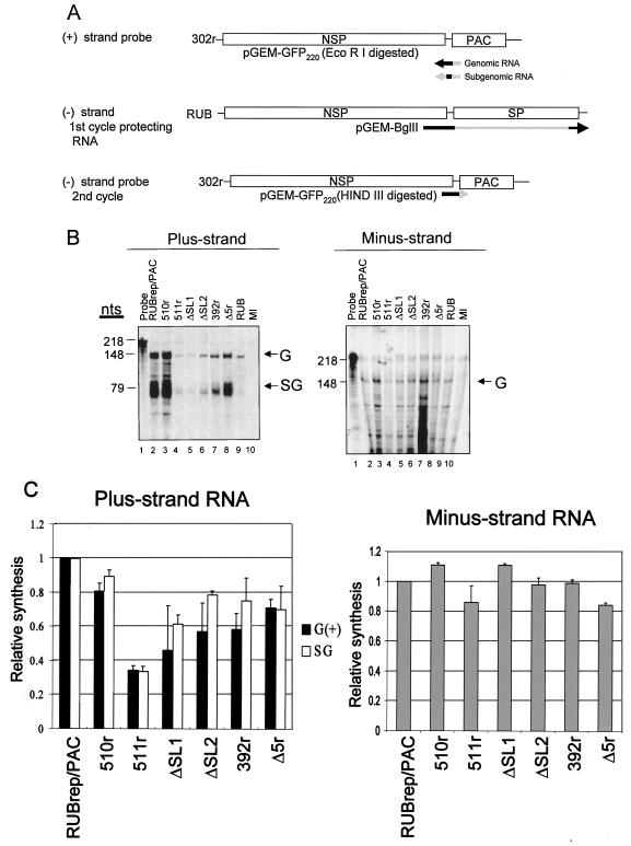
A rubella virus (RUB) replicon, RUBrep/PAC, was constructed and used to map the 3' cis-acting elements (3' CSE) of the RUB genome required for RUB replication. The RUBrep/PAC replicon had the structural protein open reading frame partially replaced by a puromycin acetyltransferase (PAC) gene. Cells transfected with RUBrep/PAC transcripts expressed the PAC gene from the subgenomic RNA, were rendered resistant to puromycin, and thus survived selection with this drug. The relative survival following puromycin selection of cells transfected with transcripts from RUBrep/PAC constructs with mutations in the 3' CSE varied. The 3' region necessary for optimal relative survival consisted of the 3' 305 nucleotides (nt), a region conserved in RUB defective-interfering RNAs, and thus this region constitutes the 3' CSE. Within the 3' CSE, deletions in the approximately 245 nt that overlap the 3' end of the E1 gene resulted in reduced relative survivals, ranging from 20 to <1% of the parental replicon survival level while most mutations within the approximately 60-nt 3' untranslated region (UTR) were lethal. None of the 3' CSE mutations affected in vitro translation of the nonstructural protein open reading frame (which is 5' proximal in the genome and encodes the enzymes involved in virus RNA replication). In cells transfected with replicons with 3' CSE mutations that survived antibiotic selection (i.e., those with mutations in the region of the 3' CSE that overlaps the E1 coding region), the amount of replicon-specific minus-strand RNA was uniform; however, the accumulation of both plus-strand RNA species, genomic and subgenomic, varied widely, indicating that this region of the RUB 3' CSE affects plus-strand RNA accumulation rather than minus-strand RNA synthesis.


Figures

FIG. 1.
FIG. 1.
Replication of the RUBrep/PAC replicon in BHK and Vero cells. (A) Schematic of RUBrep/PAC compared to RUB genome. RUBrep/PAC has most of SP-ORF replaced by a puromycin resistance (PAC) gene. As indicated, the PAC gene was preceded by an introduced sequence (in lowercase type) that included an XbaI restriction site (underlined), a TAG stop codon in frame with NS-ORF, a Kozak sequence for optimal translation initiation (in italics), and the start codon for the PAC gene. (B) Replication of RUBrep/PAC in BHK and Vero cells by Northern hybridization. Either BHK or Vero cells were transfected with RUBrep/PAC using Lipofectamine 2000 and subjected to puromycin selection, and intracellular RNA from the surviving cells at passage 3 was analyzed by Northern hybridization using a DIG-11-dUTP-labeled, nick-translated, RUBrep/PAC DNA probe. Intracellular RNA preparations from mock-infected and RUB-infected BHK or Vero cells were included for comparison. Viral genomic RNA (vG), viral subgenomic RNA (vSG), rG, and rSG are indicated.
FIG. 2.
FIG. 2.
Effects of deletions and point mutations in the 3′-terminal 600 nt of RUBrep/PAC on the generation of puromycin-resistant colonies. (A) Deletion analysis in E1 coding region. Four mutations deleting large regions of the 3′ 600 nt of the RUB genome contained in RUBrep/PAC were made as indicated. The regions of predicted RNA secondary structures in the 3′-terminal 240 nt of RUB genome are represented by different patterns of boxes (SL1 [cross-hatch pattern], SL2 [dark shading], SL3 [light shading], and SL4 [black with white dots]), while the regions without significant predicted RNA secondary structure are denoted by very light shading (white boxes with widely spaced black dots). Deletions are represented by dashed lines, and the nucleotides are numbered according to their positions in the RUB genome. Coding sequences for the reporter gene, PAC, are represented as a solid box. BHK cells were electroporated with in vitro transcripts from RUBrep/PAC or one of the RUBrep/PAC mutants. Following antibiotic selection for 7 days, the number of cells surviving antibiotic selection was determined and compared to surviving cells of RUBrep/PAC (relative survival). The relative survival shown is the average of five electroporations with each construct. Approximately 1/30 of the surviving cells from one electroporation was replated, colonies were allowed to grow, and colonies were stained in order to give a direct visualization of the number of cells surviving antibiotic selection (reverse plaque assay). (B) Mutagenic analysis of SL2 and 3′ UTR. The relative survival of various SL2 and 3′ UTR modifications of RUBrep/PAC is compared with previously published results of the same modifications in an infectious clone (Robo302) (4). The names of the RUBrep/PAC and the infectious clone construct (in parentheses) are both given (first column). A description of the construct is given (column 2); negative numbers indicate the position from the 3′ end of the RUB genome. Results from the previous work (columns 3 and 4) and the present study (column 5) with replicons are shown. Replicon results were determined as described for panel A. Relative survival (R.S.) for each replicon construct is shown. Note that the relative infectivity of the infectious clone construct 392 refers to the original mutant, not the revertant.
FIG. 2.
FIG. 2.
Effects of deletions and point mutations in the 3′-terminal 600 nt of RUBrep/PAC on the generation of puromycin-resistant colonies. (A) Deletion analysis in E1 coding region. Four mutations deleting large regions of the 3′ 600 nt of the RUB genome contained in RUBrep/PAC were made as indicated. The regions of predicted RNA secondary structures in the 3′-terminal 240 nt of RUB genome are represented by different patterns of boxes (SL1 [cross-hatch pattern], SL2 [dark shading], SL3 [light shading], and SL4 [black with white dots]), while the regions without significant predicted RNA secondary structure are denoted by very light shading (white boxes with widely spaced black dots). Deletions are represented by dashed lines, and the nucleotides are numbered according to their positions in the RUB genome. Coding sequences for the reporter gene, PAC, are represented as a solid box. BHK cells were electroporated with in vitro transcripts from RUBrep/PAC or one of the RUBrep/PAC mutants. Following antibiotic selection for 7 days, the number of cells surviving antibiotic selection was determined and compared to surviving cells of RUBrep/PAC (relative survival). The relative survival shown is the average of five electroporations with each construct. Approximately 1/30 of the surviving cells from one electroporation was replated, colonies were allowed to grow, and colonies were stained in order to give a direct visualization of the number of cells surviving antibiotic selection (reverse plaque assay). (B) Mutagenic analysis of SL2 and 3′ UTR. The relative survival of various SL2 and 3′ UTR modifications of RUBrep/PAC is compared with previously published results of the same modifications in an infectious clone (Robo302) (4). The names of the RUBrep/PAC and the infectious clone construct (in parentheses) are both given (first column). A description of the construct is given (column 2); negative numbers indicate the position from the 3′ end of the RUB genome. Results from the previous work (columns 3 and 4) and the present study (column 5) with replicons are shown. Replicon results were determined as described for panel A. Relative survival (R.S.) for each replicon construct is shown. Note that the relative infectivity of the infectious clone construct 392 refers to the original mutant, not the revertant.
FIG. 3.
FIG. 3.
Effects of 3′ modifications on NSP synthesis in replicons. Equal amounts of in vitro transcripts from the indicated RUBrep/PAC constructs were translated in a rabbit reticulocyte lysate containing [35S]methionine. Following resolution by sodium dodecyl sulfate-polyacrylamide gel electrophoresis, P200 polyprotein was quantitated by densitometry and the relative intensity, averaged from three independent experiments, was calculated by normalization to that of RUBrep/PAC. Error bars, standard deviations.
FIG. 4.
FIG. 4.
Strand-specific RNA synthesis in BHK cells transfected with various RUBrep/PAC constructs. (A) Schematic representation of probes and protecting RNA used for RPA. To detect plus-strand RNAs, including genomic and subgenomic RNAs, 35S-labeled RNA transcripts of negative polarity with a total of 218 nt were prepared by runoff transcription in vitro using digested pGEM-GFP220 and were used directly in RPA. To detect minus-strand RNA, unlabeled RNA probe transcribed from a construct, pGEM-BglII, encoding 3′ 4,000 nt in the RUB genome, was first used, and the protected products from this first cycle of RPA were hybridized to a 35S-labeled pGEM-GFP220 probe of positive polarity. The regions that would hybridize to replicon genomes are denoted as black lines, while those that would be digested from protecting RNA and probes by RNase are shown in gray. Equal amounts of RNA extracted from cells within five passages following puromycin were used for RPA analysis. (B) Representative gels are shown: left panel, plus strand RNA; right panel, minus strand RNA. The product protected by genomic (G) RNA (both plus and minus strand) was 148 nt, and that protected by the subgenomic RNA (SG) was 79 nt, as indicated on the right margin of the gel. RPA band intensities were quantitated using a FluorChem imaging system. (C) The results of quantitation are the means of three RPA analyses using intracellular RNA from two independent transfections.

References

    1. Agapov, E. V., I. Frolov, B. D. Lindenbach, B. M. Pragai, S. Schlesinger, and C. M. Rice. 1998. Noncytopathic Sindbis virus RNA vectors for heterologous gene expression. Proc. Natl. Acad. Sci. USA 95:12989-12994. - PMC - PubMed
    1. Ausubel, F. M., R. Brent., R. E. Kingston, D. D. Moore, J. G. Seidman, J. A. Smith, and K. Struhl (ed.). 1998. Current protocols in molecular biology. John Wiley and Sons, New York, N.Y.
    1. Boccard, F., and D. Baulcombe. 1993. Mutational analysis of cis-acting sequences and gene function in RNA3 of cucumber mosaic virus. Virology 193:563-578. - PubMed
    1. Chen, M.-H., and T. K. Frey. 1999. Mutagenic analysis of the 3′ cis-acting elements of the rubella virus genome. J. Virol. 73:3386-3403. - PMC - PubMed
    1. Decker, C. J., and R. Parker. 1995. Diversity of cytoplasmic functions for the 3′ untranslated region of eukaryotic transcripts. Curr. Opin. Cell Biol. 7:386-392. - PubMed

Publication types

MeSH terms