. 2004 Feb 13;32(3):1143-53.
doi: 10.1093/nar/gkh267. Print 2004.

Psoralen interstrand cross-link repair is specifically altered by an adjacent triple-stranded structure

Affiliations

Psoralen interstrand cross-link repair is specifically altered by an adjacent triple-stranded structure

F Guillonneau et al. Nucleic Acids Res. .

Abstract

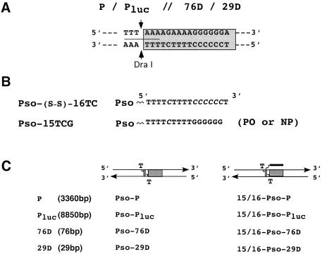
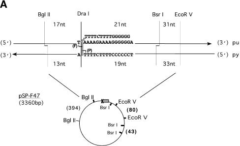
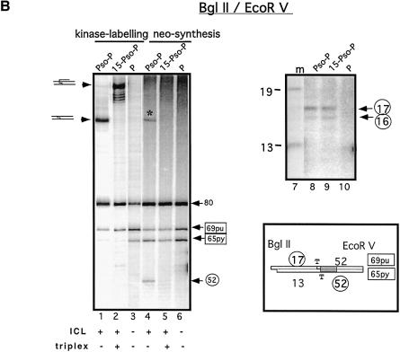
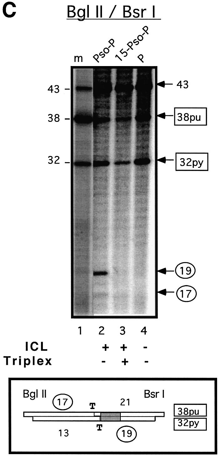
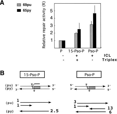
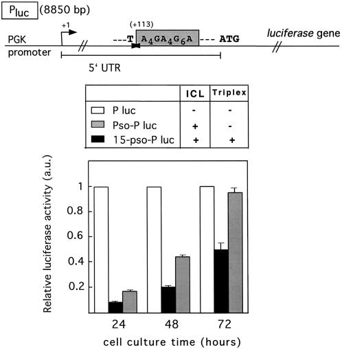
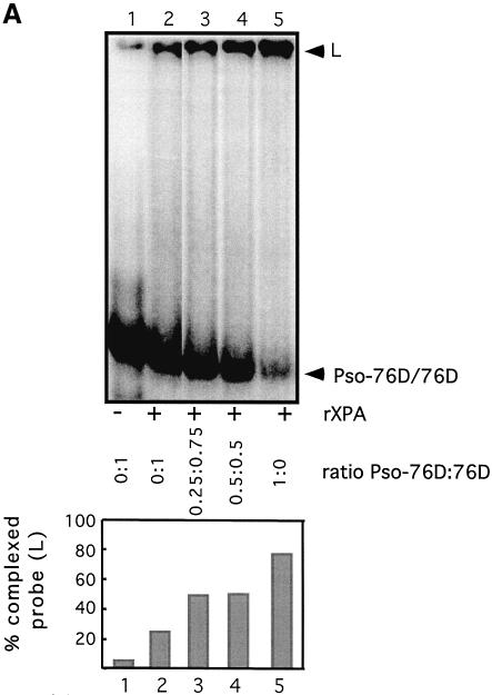
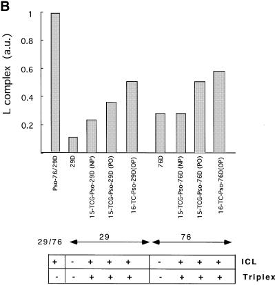
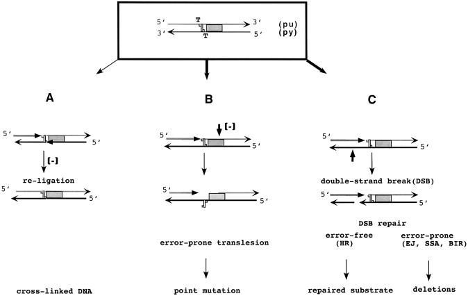
Targeting DNA-damaging agents to specific DNA sites by using sequence-specific DNA ligands has been successful in directing genomic modifications. The understanding of repair processing of such targeted damage and the influence of the adjacent complex is largely unknown. In this way, directed interstrand cross-links (ICLs) have already been generated by psoralen targeting. The mechanisms responsible for ICL removal are far from being understood in mammalian cells, with the proposed involvement of both mutagenic and recombinogenic pathways. Here, a unique ICL was introduced at a selected site by photoactivation of a psoralen moiety with the use of psoralen conjugates of triplex-forming oligonucleotides. The processing of psoralen ICL was evaluated in vitro and in cells for two types of cross-linked substrates, either containing a psoralen ICL alone or with an adjacent triple-stranded structure. We show that the presence of a neighbouring triplex structure interferes with different stages of psoralen ICL processing: (i) the ICL-induced DNA repair synthesis in HeLa cell extracts is inhibited by the triplex structure, as measured by the efficiency of 'true' and futile repair synthesis, stopping at the ICL site; (ii) in HeLa cells, the ICL removal via a nucleotide excision repair (NER) pathway is delayed in the presence of a neighbouring triplex; and (iii) the binding to ICL of recombinant xeroderma pigmentosum A protein, which is involved in pre-incision recruitment of NER factors is impaired by the presence of the third DNA strand. These data characterize triplex-induced modulation of ICL repair pathways at specific steps, which might have implications for the controlled induction of targeted genomic modifications and for the associated cellular responses.


Figures

Figure 1
Figure 1
Experimental system. (A) DNA substrates used in this study: two plasmids [pSPF47 abbreviated as P, 3360 bp long (see Fig. 2 for details), and PGK/luc abbreviated as Pluc, 8850 bp (see Fig. 4 for details)] and two synthetic double helices (76 and 29 bp long), abbreviated as 76D and 29D, respectively (for sequences, see Materials and Methods). They all contain the oligopyrimidine·oligopurine sequence suitable for triplex formation (called HIV-PPT) (grey box). A psoralen ICL can be formed at the 5′ TA 3′ site coinciding with the DraI restriction site (5′ TTT|AAA 3′, underlined). (B) The third strand oligonucleotides are represented. C stands for 5-methylated cytosines. The psoralen molecule was attached to the phosphodiester 16TC TFO via a hexamethylene arm, either directly (Pso-16TC) or via an additional disulfide bond (Pso-S-S-16TC). The Pso-15TCG oligonucleotide was used with either a phosphodiester (PO) or phosphoramidate (NP) backbone. (C) The two types of substrates containing a psoralen ICL at the 5′ TA 3′ site used in this study are schematically represented. Left: DNA substrates with a unique psoralen ICL, obtained after Pso-S-S-16TC (cleavable) treatment (Pso-P, Pso-Pluc and Pso-76D, Pso-29D). Right: DNA substrates with a unique psoralen ICL and an adjacent triplex structure obtained after Pso-16TC or Pso-15TCG (uncleavable) treatment (16 or 15-Pso-P, 16 or 15-Pso-Pluc, and 16 or 15-Pso-76D, 16 or 15-Pso-29D) (for details see Materials and Methods). The triplex site is shown (grey box). The total lengths of the various DNA substrates are indicated.
Figure 2
Figure 2
Analysis of products obtained by repair processing of psoralen cross-linked substrates in HeLa cell extracts. Synthesis repair assays were performed (as described in Materials and Methods) and, after purification, DNA fragments were digested with the indicated restriction enzymes and revealed by denaturing PAGE. (A) Schematic representation of the pSP-F47 plasmid (P) in the region surrounding the defined psoralen ICL site. Cleavage sites of the restriction enzymes on both DNA strands are indicated by vertical bars. The numbers between the bars indicate the distances in nucleotides between the cleavage sites. The thymines that are cross-linked in the psoralen-modified substrates, Pso-P and 15-Pso-P, are indicated. The orientation of the psoralen molecule is described with the furane (F) and the pyrone (P) sides. A schematic map of the pSP-F47 plasmid is represented with the triplex site indicated by a box and the ICL site by a cross (X). The 80 or 43 fragments are unmodified fragments used as references using BglII–EcoRV or BglII–BsrI digestion, respectively. Fragments sizes are indicated. (B) Analysis of species after BglII–EcoRV digestion. Lanes 1–3: for the three plasmids, unmodified (P) or containing a unique psoralen ICL (Pso-P and 15-Pso-P), digestion fragments were 5′ labelled (see Materials and Methods). Lanes 4–6 and 8–10: neo-synthesized DNA fragments obtained upon incubation with HeLa cell extracts. 69pu and 65py fragments correspond to full-length repair products, and contain the oligopyrimidine·oligopurine PPT sequence. The 52 and 17/16 nt fragments correspond to futile repair products stopping at the ICL site (pyrimidine and purine strand, respectively). The 80 nt fragments did not contain the ICL site and PPT target sequence (see A) and were used as references for quantification of relative repair activity (see Fig. 3A and Materials and Methods). Cross-linked species are indicated on the left of the gel. The asterisk indicates the fragments of neo-synthesis, religated to the cross-linked thymine (see text for details). Lane 7 is a size marker obtained by 5′ labelling of digestion products; DraI digestions of neo-synthesized 65py and 69pu fragments were indistinguishable from futile synthesis products (52 and 17 nt fragments) (data not shown). (C) Analysis of neo-synthesized species after BglII and BsrI digestion for various plasmid templates, as indicated. Full-length (38pu and 32py) and futile repair (17 and 19) fragments are indicated. The 17 nt fragment is hardly detected (see stoichiometries in Fig. 3B). The 43 nt fragments were used as reference fragments since they do not contain the target sequence (see A). Lane 1 is a size marker obtained by 5′ labelling of BglII/BsrI/DraI digestion products. A schematic representation of the substrates is given below (B) and (C): the triplex site is boxed, the adducted thymines are indicated and the various fragments are depicted with their respective lengths.
Figure 2
Figure 2
Analysis of products obtained by repair processing of psoralen cross-linked substrates in HeLa cell extracts. Synthesis repair assays were performed (as described in Materials and Methods) and, after purification, DNA fragments were digested with the indicated restriction enzymes and revealed by denaturing PAGE. (A) Schematic representation of the pSP-F47 plasmid (P) in the region surrounding the defined psoralen ICL site. Cleavage sites of the restriction enzymes on both DNA strands are indicated by vertical bars. The numbers between the bars indicate the distances in nucleotides between the cleavage sites. The thymines that are cross-linked in the psoralen-modified substrates, Pso-P and 15-Pso-P, are indicated. The orientation of the psoralen molecule is described with the furane (F) and the pyrone (P) sides. A schematic map of the pSP-F47 plasmid is represented with the triplex site indicated by a box and the ICL site by a cross (X). The 80 or 43 fragments are unmodified fragments used as references using BglII–EcoRV or BglII–BsrI digestion, respectively. Fragments sizes are indicated. (B) Analysis of species after BglII–EcoRV digestion. Lanes 1–3: for the three plasmids, unmodified (P) or containing a unique psoralen ICL (Pso-P and 15-Pso-P), digestion fragments were 5′ labelled (see Materials and Methods). Lanes 4–6 and 8–10: neo-synthesized DNA fragments obtained upon incubation with HeLa cell extracts. 69pu and 65py fragments correspond to full-length repair products, and contain the oligopyrimidine·oligopurine PPT sequence. The 52 and 17/16 nt fragments correspond to futile repair products stopping at the ICL site (pyrimidine and purine strand, respectively). The 80 nt fragments did not contain the ICL site and PPT target sequence (see A) and were used as references for quantification of relative repair activity (see Fig. 3A and Materials and Methods). Cross-linked species are indicated on the left of the gel. The asterisk indicates the fragments of neo-synthesis, religated to the cross-linked thymine (see text for details). Lane 7 is a size marker obtained by 5′ labelling of digestion products; DraI digestions of neo-synthesized 65py and 69pu fragments were indistinguishable from futile synthesis products (52 and 17 nt fragments) (data not shown). (C) Analysis of neo-synthesized species after BglII and BsrI digestion for various plasmid templates, as indicated. Full-length (38pu and 32py) and futile repair (17 and 19) fragments are indicated. The 17 nt fragment is hardly detected (see stoichiometries in Fig. 3B). The 43 nt fragments were used as reference fragments since they do not contain the target sequence (see A). Lane 1 is a size marker obtained by 5′ labelling of BglII/BsrI/DraI digestion products. A schematic representation of the substrates is given below (B) and (C): the triplex site is boxed, the adducted thymines are indicated and the various fragments are depicted with their respective lengths.
Figure 3
Figure 3
Quantification of the abundance of the neo-synthesized fragments due to repair processing of the different cross-linked DNA substrates. (A) Relative repair activities (R) of full-length repair products: purine (light grey) and pyrimidine (dark grey) strands were reported for each template (as indicated). They were obtained after dividing the relative abundance of neo-synthesized products (obtained by nucleotide incorporation) by their relative abundance in the initial mixture (obtained by kinase labelling) (average of three experiments; see details in Materials and Methods). (B) Schematic representation of the various neo-synthesis fragments obtained in vitro for the two different psoralen cross-linked substrates used in the present work. Purine and pyrimidine full-length or futile repair fragments are represented with their associated stoichiometries, calculated on the basis of fragment signal intensities and cytosine contents (mean from four experiments). For each of the full-length fragments, the background synthesis, estimated from (A) by comparison of the undamaged substrate (P) with the damaged ones (Pso-P and 15-Pso-P), was deduced in order to obtain the level of repair-induced synthesis.
Figure 4
Figure 4
Kinetics of cross-link repair in cells. The PGK/luc plasmid (Pluc) expressing the firefly luciferase was modified in order to obtain a single cross-linked triplex (15-Pso-Pluc) or a unique psoralen ICL (Pso-Pluc) in the 5′-untranslated region (see Materials and Methods). Luciferase activities were measured at different times after transfection (24, 48 and 72 h) in HeLa cells. In all experiments, plasmids were co-transfected with a control vector expressing Renilla luciferase. Reporter activity is presented as the normalized ratio between firefly and Renilla luciferase activities in the same lysate (relative luciferase activity). Normalization from cells transfected with an unmodified plasmid P permits quantification of the cross-link repair efficiency. The error bars correspond to the mean deviation from duplicate experiments.
Figure 5
Figure 5
XPA binding to various types of psoralen ICL. (A) Gel retardation analysis of rXPA binding to the 76D duplex containing a unique psoralen ICL (Pso-6D) in various proportions, as indicated by the ratio Pso-76D:76D. The radiolabelled duplex concentration (Pso-76D + 76D) is 10 nM, and rXPA protein (1 µM) was added or not, as indicated. The amount of retarded complex L is indicated below the gel. (B) Triplex interferes with XPA binding to psoralen-modified duplexes. The damaged duplexes (16-Pso-76 or 29D and Pso-76 or 29D) were used as probes; complex formation was achieved by addition of rXPA (1 µM). Amounts of L complex (a.u.) obtained with various psoralen-damaged probes are reported; the values are normalized to that obtained for Pso-76/29D substrates. Values from one representative experiment that was independently repeated twice are presented. Equivalent results were obtained with the 76D and the 29D substrates.
Figure 5
Figure 5
XPA binding to various types of psoralen ICL. (A) Gel retardation analysis of rXPA binding to the 76D duplex containing a unique psoralen ICL (Pso-6D) in various proportions, as indicated by the ratio Pso-76D:76D. The radiolabelled duplex concentration (Pso-76D + 76D) is 10 nM, and rXPA protein (1 µM) was added or not, as indicated. The amount of retarded complex L is indicated below the gel. (B) Triplex interferes with XPA binding to psoralen-modified duplexes. The damaged duplexes (16-Pso-76 or 29D and Pso-76 or 29D) were used as probes; complex formation was achieved by addition of rXPA (1 µM). Amounts of L complex (a.u.) obtained with various psoralen-damaged probes are reported; the values are normalized to that obtained for Pso-76/29D substrates. Values from one representative experiment that was independently repeated twice are presented. Equivalent results were obtained with the 76D and the 29D substrates.
Figure 6
Figure 6
Model for repair of psoralen ICL and modulation by an adjacent triplex structure, based on the presented findings. Three possible repair pathways are described with futile synthesis repair fragments (grey arrows) as a starting point. (A) Following filling in, a small fraction of molecules are ligated to regenerate a cross-linked substrate. (B) 3′ Cleavage (indicated by a vertical arrow) occurring on the same strand as the neo-synthesized fragment produces a gap which can be replenished by translesion followed by ligation and, probably, an additional cleavage process to completely remove the damage. This pathway is mutagenic. (C) 3′ Cleavage (indicated by a vertical arrow) occurring on the strand opposite to the neo-synthesized product produces a DSB that can be repaired by either error-free HR or error-prone EJ, SSA and BIR processes (see text for detailed discussion). The different stages where the triplex was shown in the present study to interfere with ICL repair are indicated by (–). It is noticeable that for the cross-linked triplex, the pathway in (A) seems to be ineffective, and the pathways in (B) and (C) can only occur for the truncated neo-synthesized purine strand since a complete blockage of futile synthesis of the pyrimidine strand is observed (see Fig. 3B). The triplex site is boxed (in grey) and the adducted thymines are indicated.

Similar articles

Cited by

References

    1. Faria M. and Giovannangeli,C. (2001) Triplex-forming molecules: from concepts to applications. J. Gene Med., 3, 299–310. - PubMed
    1. Knauert M.P. and Glazer,P.M. (2001) Triplex forming oligonucleotides: sequence-specific tools for gene targeting. Hum. Mol. Genet., 10, 2243–2251. - PubMed
    1. Seidman M.M. and Glazer,P.M. (2003) The potential for gene repair via triple helix formation. J. Clin. Invest., 112, 487–494. - PMC - PubMed
    1. De Silva I.U., McHugh,P.J., Clingen,P.H. and Hartley,J.A. (2000) Defining the roles of nucleotide excision repair and recombination in the repair of DNA interstrand cross-links in mammalian cells. Mol. Cell. Biol., 20, 7980–7990. - PMC - PubMed
    1. Wang G., Chen,Z., Zhang,S., Wilson,G.L. and Jing,K. (2001) Detection and determination of oligonucleotide triplex formation-mediated transcription-coupled DNA repair in HeLa nuclear extracts. Nucleic Acids Res., 29, 1801–1807. - PMC - PubMed

Publication types