. 2004 Mar;24(5):1844-54.
doi: 10.1128/MCB.24.5.1844-1854.2004.

The NAD(P)H oxidase homolog Nox4 modulates insulin-stimulated generation of H2O2 and plays an integral role in insulin signal transduction

Affiliations

The NAD(P)H oxidase homolog Nox4 modulates insulin-stimulated generation of H2O2 and plays an integral role in insulin signal transduction

Kalyankar Mahadev et al. Mol Cell Biol. 2004 Mar.

Abstract

Insulin stimulation of target cells elicits a burst of H(2)O(2) that enhances tyrosine phosphorylation of the insulin receptor and its cellular substrate proteins as well as distal signaling events in the insulin action cascade. The molecular mechanism coupling the insulin receptor with the cellular oxidant-generating apparatus has not been elucidated. Using reverse transcription-PCR and Northern blot analyses, we found that Nox4, a homolog of gp91phox, the phagocytic NAD(P)H oxidase catalytic subunit, is prominently expressed in insulin-sensitive adipose cells. Adenovirus-mediated expression of Nox4 deletion constructs lacking NAD(P)H or FAD/NAD(P)H cofactor binding domains acted in a dominant-negative fashion in differentiated 3T3-L1 adipocytes and attenuated insulin-stimulated H(2)O(2) generation, insulin receptor (IR) and IRS-1 tyrosine phosphorylation, activation of downstream serine kinases, and glucose uptake. Transfection of specific small interfering RNA oligonucleotides reduced Nox4 protein abundance and also inhibited the insulin signaling cascade. Overexpression of Nox4 also significantly reversed the inhibition of insulin-stimulated IR tyrosine phosphorylation induced by coexpression of PTP1B by inhibiting PTP1B catalytic activity. These data suggest that Nox4 provides a novel link between the IR and the generation of cellular reactive oxygen species that enhance insulin signal transduction, at least in part via the oxidative inhibition of cellular protein-tyrosine phosphatases (PTPases), including PTP1B, a PTPase that has been previously implicated in the regulation of insulin action.


Figures

FIG. 1.
FIG. 1.
Schematic structure of Nox4 wild-type and deletion constructs expressed in 3T3-L1 cells by adenoviral gene delivery. The full-length wild-type Nox4 protein is shown with the major functional domains indicated. The construct with the deleted NAD(P)H binding domain is designated ΔNADPH, and the construct with both cofactor domains deleted is indicated as ΔFAD/NADPH.
FIG. 2.
FIG. 2.
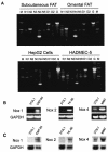
Expression of Nox family homologs in adipose tissue and various cultured cells. (A) RT-PCR of Nox family homologs. Total RNA was prepared using the TRIzol reagent, and RT-PCR was performed with primers specific for human Nox1, Nox2, Nox3, Nox4, Nox5, Duox1, and Duox2 in human subcutaneous and omental adipose tissue, HepG2 hepatoma cells, and SV40-transformed human microvascular endothelial (HADMEC-5) cells using previously reported techniques (8). N1 to N5, Nox1 to Nox5; D1 and D2, Duox 1 and 2; G, glyceraldehyde-3-phosphate dehydrogenase (GAPDH) as an internal control; M, molecular size markers. (B) RT-PCR was performed using RNA isolated from differentiated 3T3-L1 adipocytes as described in Materials and Methods using primers for PCR analysis that were specific to murine Nox family homologs Nox1, Nox2, and Nox4. Control cell lines included CMT-93 (murine colon carcinoma cells), HL-60 (human leukemia cells), and MMC (murine mesangial cells). (C) Northern blot analysis of Nox4 mRNA expression in differentiated 3T3-L1 adipocytes. Total RNA was isolated from the indicated cells using the TRIzol reagent, and Northern blot analysis using Nox1-, Nox2-, and Nox4-specific cDNA probes or GAPDH as a loading and transfer control was performed as described in Materials and Methods.
FIG. 3.
FIG. 3.
Expression of Nox4 protein in 3T3-L1 adipocytes after transfection of siRNA oligonucleotides and expression of recombinant Nox4 mRNA and PTP1B protein in 3T3-L1 adipocytes following adenovirus-mediated gene delivery. (A) 3T3-L1 adipocytes were transfected with Nox4-specific (siRNA 1, 3, or both together) or a scrambled control siRNA oligonucleotide and reseeded in six-well plates (6). After 48 h of incubation, cell lysates were prepared and incubated with Nox4 polyclonal antibody to immunoprecipitate Nox4 protein. Immunoprecipitates were separated by SDS-PAGE and reblotted with Nox4 antibody as described in Materials and Methods. A representative blot demonstrating expression of Nox4 protein at ∼65 kDa and a reduction in Nox4 abundance following loading of specific siRNA oligonucleotides is shown. Protein expression for the insulin receptor β-subunit is shown as an experimental control. The bar graph shows mean data from replicate immunoblots quantitated using ImageStation 440 (Kodak). *, P = 0.05; **, P = 0.01 versus control cells transfected with the scrambled siRNA. (B) At 72 h following transduction of differentiated 3T3-L1 adipocytes with adenovirus encoding β-galactosidase or the indicated Nox4 constructs, total RNA was prepared using the TRIzol reagent and RT-PCR was performed with Nox4-specific primers as described in Materials and Methods. Nox4 was detected as a ∼550-bp band in the ethidium bromide-stained agarose gel. (C) Cell lysates were prepared from 3T3-L1 cells 72 h after transduction with control adenovirus encoding β-galactosidase, wild-type PTP1B, or a site-directed catalytically inactive (Cys215S→er) PTP1B construct (mPTP1B). Proteins were separated by SDS-PAGE and immunoblotted with PTP1B antibody as described in Materials and Methods. The 50-kDa PTP1B protein band is indicated. Protein expression for the insulin receptor β-subunit is shown as an experimental control.
FIG. 4.
FIG. 4.
Effect of adenoviral expression of recombinant Nox4 and PTP1B constructs on insulin-stimulated production of H2O2 in 3T3-L1 adipocytes. Seventy-two hours after transduction with the indicated adenovirus constructs, differentiated 3T3-L1 adipocytes were starved overnight in serum-free medium, loaded with CM-DCF-DA, and stimulated with 100 nM insulin for 5 min as described in Materials and Methods. Intracellular H2O2 production was detected by fluorescence of DCF in situ using confocal microscopy as reported previously (35). *, P = 0.02; ***, P < 0.001 versus control.
FIG. 5.
FIG. 5.
Effect of adenoviral expression of recombinant Nox4 and PTP1B constructs on insulin-stimulated tyrosine phosphorylation of the insulin receptor β-subunit and IRS proteins. (A) Representative antiphosphotyrosine (4G10) and anti-insulin receptor β-subunit immunoblots of 3T3-L1 cell lysates transduced with adenovirus encoding wild-type Nox4 as well as two dominant-negative constructs lacking binding domains for NAD(P)H or FAD/NAD(P)H or wild-type PTP1B and mutant PTP1B, following stimulation of serum-starved cells with 100 nM insulin for 5 min. The migration positions of the tyrosine-phosphorylated insulin receptor β-subunit (95 kDa) are indicated as pY-IR-β. Mean data for the phosphotyrosine density of the insulin receptor β-subunit from replicate immunoblots quantitated using an ImageStation 440 (Kodak) are shown in the bar graph. ***, P ≤ 0.001 versus control β-galactosidase-encoding adenovirus-transduced 3T3-L1 adipocytes. (B) Samples identical to those described in the legend for panel A were analyzed for phosphotyrosine content of IRS1/2 and IRS1 protein using antiphosphotyrosine antibodies (4G10) and IRS-1 antibody following stimulation of serum-starved cells with 100 nM insulin for 5 min. The migration positions of the tyrosine-phosphorylated IRS1/2 (∼ 185 kDa) are indicated as pY IRS-1/2. Mean data for the phosphotyrosine density of IRS-1/2 from replicate immunoblots are shown in the bar graph. **, P = 0.01; ***, P ≤ 0.001 versus control cells transduced with the β-galactosidase-encoding adenovirus. (C) Representative antiphosphotyrosine (4G10) and anti-insulin receptor β-subunit immunoblots of 3T3-L1 cell lysates transduced with adenovirus encoding wild-type PTP1B or mutant PTP1B and cotransduced with adenovirus encoding Nox4 or the FAD/NAD(P)H Nox4 deletion construct where indicated, following stimulation of serum-starved cells with 100 nM insulin for 5 min. Mean data for the phosphotyrosine density of the insulin receptor β-subunit from replicate immunoblots are shown in the bar graph. **, P = 0.01; ***, P ≤ 0.001 versus control cells transduced with the β-galactosidase-encoding adenovirus. (D) Effect of reduction of Nox4 mass on insulin-stimulated tyrosine phosphorylation of the insulin receptor β-subunit, as shown in representative antiphosphotyrosine (4G10) and anti-insulin receptor β-subunit (IR-β) immunoblots of 3T3-L1 cell lysates transfected with control (scrambled) or Nox4-specific siRNA oligonucleotides. Following stimulation of serum-starved cells with 100 nM insulin for 5 min, cell lysates were prepared and SDS-PAGE and immunoblotting were performed. Mean data for the phosphotyrosine density of the insulin receptor β-subunit from replicate immunoblots are shown in the bar graph. *, P = 0.05; **, P = 0.01 versus control transfected 3T3-L1 adipocytes.
FIG. 6.
FIG. 6.
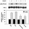
Effect of adenoviral expression of Nox4 deletion constructs and wild-type PTP1B on the association of the p85 subunit of PI 3′-kinase with IRS-1. Transduction of 3T3-L1 adipocytes with adenovirus, stimulation of cells with insulin, and preparation of lysates were performed as described in the legend to Fig. 4. After immunoprecipitation of IRS-1 from the lysates as described in Materials and Methods, samples were separated by gel electrophoresis, transferred to PVDF membranes, and probed overnight with antibody to the p85 noncatalytic subunit of PI 3′-kinase. After stripping, PVDF membranes were reprobed with the IRS-1 antibody for normalization. The upper panel shows a representative immunoblot, and the bar graph in the lower panel represents the p85 subunit binding to IRS-1 protein from replicate immunoblots after quantitation. **, P < 0.01; ***, P < 0.001 versus control β-galactosidase-encoding adenovirus-transduced 3T3-L1 adipocytes.
FIG. 7.
FIG. 7.
Effect of siRNA-induced reduction of Nox4 protein mass on insulin-stimulated Akt phosphorylation in 3T3-L1 adipocytes. Results shown are representative anti-phospho-Akt and anti-Akt protein immunoblots of 3T3-L1 cell lysates from cells transfected with control (scrambled) or Nox4-specific siRNA oligonucleotides and following stimulation of serum-starved cells with 100 nM insulin for 5 min as described in Materials and Methods. The bar graph represents mean data from replicate immunoblots after quantitation of signal density. *, P = 0.05; **, P = 0.01; ***, P = 0.007 versus control 3T3-L1 adipocytes transfected with the scrambled siRNA.
FIG. 8.
FIG. 8.
Effect of adenoviral expression of recombinant Nox4 and PTP1B constructs on Erk1 and Erk2 MAPK activation in 3T3-L1 adipocytes. Representative anti-phospho-MAPK and anti-MAPK protein immunoblots of lysates from 3T3-L1 cells transduced with the indicated adenoviral constructs are shown. Following stimulation of serum-starved cells with 100 nM insulin for 5 min, cell lysates were prepared and immunoblotting was performed with a monoclonal antibody to detect phospho-MAPK (ERK1/2) or anti-MAPK protein antibody. (Upper panel) Representative immunoblots demonstrating the phosphorylation state of MAPK and the MAPK protein level. (Lower panel) Mean data from replicate immunoblots as shown in the upper panel after quantitation of signal density. *, P = 0.05 versus control β-galactosidase-encoding adenovirus-transduced 3T3-L1 adipocytes.
FIG. 9.
FIG. 9.
Effect of adenoviral expression of recombinant Nox4 or PTP1B or siRNA-induced reduction in Nox4 protein mass on insulin-stimulated glucose uptake in 3T3-L1 adipocytes. (A) After differentiation, 3T3-L1 adipocytes were transduced with adenovirus encoding β-galactosidase as a control, wild-type Nox4, or constructs lacking binding domains for NAD(P)H or FAD/NAD(P)H as shown and serum starved for 2 h prior to stimulation with 100 nM insulin and measurement of glucose transport by the uptake of 2-deoxy-d-glucose as described in Materials and Methods. (B) Glucose transport measured in differentiated 3T3-L1 cells after transfection with control (scrambled) or Nox4-specific siRNA oligonucleotides as shown. *, P < 0.05; **, P < 0.01; ***, P < 0.001 versus control cells stimulated with insulin.
FIG. 10.
FIG. 10.
Effect of adenoviral expression of recombinant PTP1B without or with Nox4 on the endogenous catalytic activity of PTP1B in 3T3-L1 adipocytes. Differentiated 3T3-L1 adipocytes were transduced with adenovirus encoding β-galactosidase as a control or wild-type PTP1B without or with cotransduction of recombinant active Nox4 as shown. Cells were serum starved for 2 h prior to stimulation with 100 nM insulin for 5 min. Cell lysates were immunoprecipitated with a specific antibody for PTP1B, and PTPase activity of the adsorbed enzyme was measured under an insert atmosphere by hydrolysis of para-nitrophenyl phosphate as described in Materials and Methods. ***, P < 0.001 versus the respective control samples without or with insulin.

References

    1. Babior, B. M., J. D. Lambeth, and W. Nauseef. 2002. The neutrophil NADPH oxidase. Arch. Biochem. Biophys. 397:342-344. - PubMed
    1. Bae, Y. S., S. W. Kang, M. S. Seo, I. C. Baines, E. Tekle, P. B. Chock, and S. G. Rhee. 1997. Epidermal growth factor (EGF)-induced generation of hydrogen peroxide. Role in EGF receptor-mediated tyrosine phosphorylation. J. Biol. Chem. 272:217-221. - PubMed
    1. Bae, Y. S., J. Y. Sung, O. S. Kim, Y. J. Kim, K. C. Hur, A. Kazlauskas, and S. G. Rhee. 2000. Platelet-derived growth factor-induced H2O2 production requires the activation of phosphatidylinositol 3-kinase. J. Biol. Chem. 275:10527-10531. - PubMed
    1. Banfi, B., R. A. Clark, K. Steger, and K. H. Krause. 2003. Two novel proteins activate superoxide generation by the NADPH oxidase NOX1. J. Biol. Chem. 278:3510-3513. - PubMed
    1. Barrett, W. C., J. P. DeGnore, Y. F. Keng, Z. Y. Zhang, M. B. Yim, and P. B. Chock. 1999. Roles of superoxide radical anion in signal transduction mediated by reversible regulation of protein-tyrosine phosphatase 1B. J. Biol. Chem. 274:34543-34546. - PubMed

Publication types

MeSH terms