. 2004 Mar;24(5):1870-83.
doi: 10.1128/MCB.24.5.1870-1883.2004.

The scl +18/19 stem cell enhancer is not required for hematopoiesis: identification of a 5' bifunctional hematopoietic-endothelial enhancer bound by Fli-1 and Elf-1

Affiliations

The scl +18/19 stem cell enhancer is not required for hematopoiesis: identification of a 5' bifunctional hematopoietic-endothelial enhancer bound by Fli-1 and Elf-1

Berthold Göttgens et al. Mol Cell Biol. 2004 Mar.

Abstract

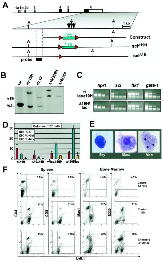
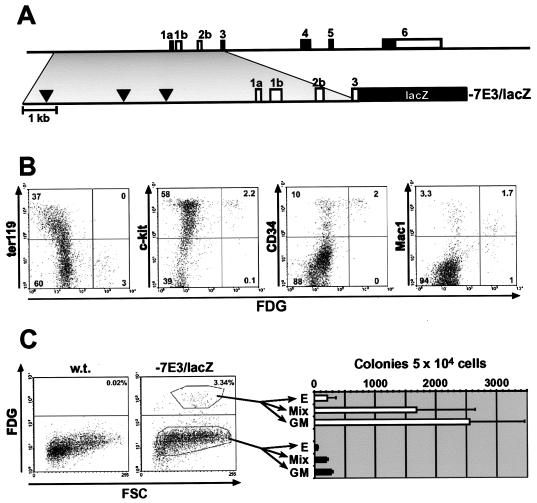
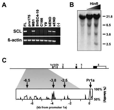
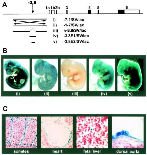
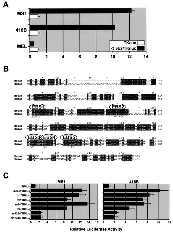
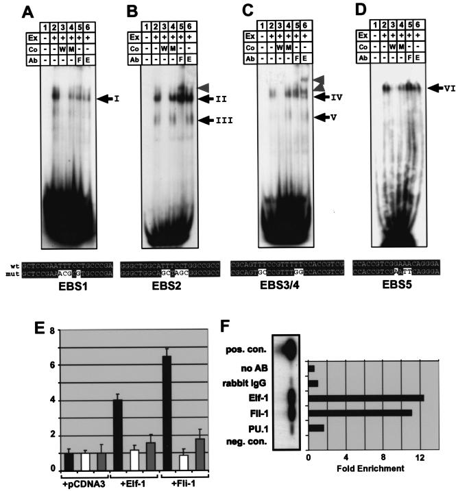
Analysis of cis-regulatory elements is central to understanding the genomic program for development. The scl/tal-1 transcription factor is essential for lineage commitment to blood cell formation and previous studies identified an scl enhancer (the +18/19 element) which was sufficient to target the vast majority of hematopoietic stem cells, together with hematopoietic progenitors and endothelium. Moreover, expression of scl under control of the +18/19 enhancer rescued blood progenitor formation in scl(-/-) embryos. However, here we demonstrate by using a knockout approach that, within the endogenous scl locus, the +18/19 enhancer is not necessary for the initiation of scl transcription or for the formation of hematopoietic cells. These results led to the identification of a bifunctional 5' enhancer (-3.8 element), which targets expression to hematopoietic progenitors and endothelium, contains conserved critical Ets sites, and is bound by Ets family transcription factors, including Fli-1 and Elf-1. These data demonstrate that two geographically distinct but functionally related enhancers regulate scl transcription in hematopoietic progenitors and endothelial cells and suggest that enhancers with dual hematopoietic-endothelial activity may represent a general strategy for regulating blood and endothelial development.


Figures

FIG.1.
FIG.1.
The scl 3′ enhancer is not required for blood formation from ES cells. (A) Strategy for deleting the mouse scl 3′ stem cell enhancer by homologous recombination in ES cells. Shown are the structure of the mouse scl locus, together with the enhancer knockout construct and the two knockout alleles (sclHΔ19 and sclΔ19) generated in the present study. Arrows indicate previously mapped DNase I-hypersensitive sites +18 and +19. (A) ApaI restriction site; hyg, hygromycin selection cassette; red arrowheads, loxP sites; probe, position of probe used for Southern analysis shown in panel B. (B) Southern blot analysis of targeted ES cell lines. DNA was digested with ApaI and analyzed by using the probe shown in panel A. Bands distinguishing the three alleles are indicated as wt (5.2 kb), HΔ19 (9.2 kb), or Δ19 (6.9 kb). (C) Levels of expression of scl, FLK1, and GATA-1 are not significantly different between embryoid bodies derived from scl+/lacHΔ19 and sclHΔ19/lac ES cells. Total RNA extracted from day 7 embryoid bodies with the indicated genotypes was reverse transcribed, and the resultant cDNA was analyzed by semiquantitative PCR. Shown are three fivefold serial dilutions and a negative control for each primer pair. (D) ES cells lacking the 18/19 enhancer can give rise to hematopoietic colonies in vitro. Cells derived from day 10 embryoid bodies of the indicated genotypes were used to perform hematopoietic colony assays. Red bars, BFU-E; turquoise bars, CFU-GM; yellow bars, CFU-Mix. The data shown are from a representative experiment. Similar results were obtained in three independent experiments. (E) Morphology of hematopoietic cells in colonies derived from sclΔ19/Δ19 ES cells (May-Grunwald-Giemsa stain). (F) sclHΔ19/lac ES cells can give rise to blood cells in vivo. FACS analysis of spleen and bone marrow cells from chimeras generated by using sclHΔ19/lac ES cells, together with control C57BL/6 and 129 mice (negative and positive controls, respectively). Ly9.1 is expressed by ES cell-derived progeny but not by host C57BL/6 cells.
FIG. 2.
FIG. 2.
The mouse scl 5′ flanking region directs expression to hematopoietic progenitor cells. (A) Diagram of mouse scl locus indicating the position of the 5′ fragment present in the −7E3/lacZ transgene. Black arrowheads indicate the positions of DNase I-hypersensitive sites previously mapped in myeloid cell lines. (B) Characterization of hematopoietic cells targeted by the 5′ enhancer. FACS analysis of E11.5 fetal liver demonstrates enhancer activity in a subset of c-kit+ cells, CD34+ cells, and Mac1+ cells, markers previously shown to be expressed by fetal liver hematopoietic stem/progenitor cells. (C) E11.5 fetal liver cells expressing the −7E3/lacZ transgene are enriched for hematopoietic progenitors. lacZ-positive and lacZ-negative cells from mice carrying the −7E3/lacZ transgene were sorted by FACS and assessed for hematopoietic colony-forming activity. FDG, fluorescein di-β-d-galactopyranoside fluorescent β-galactosidase/lacZ substrate; FSC, forward scatter; E, burst-forming units; GM, granulocyte/macrophage colonies; Mix, multipotent colonies.
FIG. 3.
FIG. 3.
Chromatin accessibility and sequence homology identify a candidate scl 5′ enhancer in endothelial cell lines. (A) RT-PCR analysis demonstrates that three of seven mouse endothelial cell lines (MS1, bEND, and sEND) express readily detectable levels of scl. FL, E14 fetal liver-positive control; (−), water-negative control. (B) Restriction endonuclease accessibility assay identifies three regions of accessible chromatin at kb −2.5, −3.8, and −6.5 upstream of mouse scl exon 1a. (C) Diagram of murine scl locus indicating 5′ regions of open chromatin (arrows) and the 6.3-kb fragment with endothelial activity in transgenic mice (shaded area). The homology profile of a mouse/human sequence alignment demonstrates that the kb −3.8 region exhibits the highest level of sequence conservation within the 6.3-kb fragment. Pr1a, promoter 1a.
FIG. 4.
FIG. 4.
The −3.8 region is both necessary and sufficient for endothelial and hematopoietic activity in transgenic mouse embryos. (A) Transgenic reporter constructs in relation to the mouse scl locus. (B) Panels i to v show representative E11.5 embryos transgenic for the constructs indicated in panel A. (C) Sections of embryo shown in panel Bv demonstrating specific expression in endothelial cells, endocardium, round cells in the fetal liver, and presumed hematopoietic clusters on the ventral wall of the dorsal aorta.
FIG. 5.
FIG. 5.
Activity of the −3.8 core enhancer requires conserved Ets transcription factor-binding sites. (A) Lineage-specific activity of the −3.8 core enhancer in endothelial and hematopoietic cell lines. Transient-transfection assays with luciferase reporter constructs show that the −3.8 core region has strong enhancer activity in MS1 (endothelial) and 416B (hematopoietic progenitor) but not MEL cells (erythroid). (B) Mouse/human sequence alignment of the −3.8 core enhancer. EBS1 to -5 indicate the positions of the four conserved TTCC/GGAA Ets binding sites. (C) Reporter assays of wild-type and mutant core enhancer constructs in MS1 and 416B cells. m1 to m5 indicate mutations in sites EBS1 to -5.
FIG. 6.
FIG. 6.
Fli-1 and Elf-1 can activate the −3.8 core enhancer and bind to it in vitro and in vivo. (A to D) Electrophoretic mobility shift assays for oligonucleotides EBS1 (A), EBS2 (B), EBS3/4 (C), and EBS5 (D) containing the Ets consensus-binding sites indicated in Fig. 5. Ex, nuclear extract; Co, competitor oligo; Ab, supershift antibody; W, wild type; M, mutant; F, Fli-1; E, Elf-1. The sequences of the wild-type oligonucleotides and the respective mutant oligonucleotides are shown underneath each panel. wt, wild-type oligonucleotide; mut, mutant oligonucleotide. (A) One major complex (complex I) was bound by the EBS1 oligonucleotide (compare lanes 1 and 2). Complex I binding was competed for by excess wild-type oligonucleotide and, to a lesser extent, by mutant oligonucleotide (compare lanes 3 and 4). Complex I was not supershifted with antibodies against Fli-1 or Elf-1 (see lanes 5 and 6). (B) Two major complexes (complexes II and III) were bound by the EBS2 oligonucleotide (compare lanes 1 and 2). Both complexes were competed by excess wild-type, but not mutant oligonucleotide (compare lanes 3 and 4). Complex II was not supershifted with antibodies against Fli-1 or Elf-1, whereas complex III was supershifted with antibody to Fli-1 but not Elf-1 (see lanes 5 and 6 and arrowhead). (C) One major complex (complex IV) was bound by the EBS3/4 oligonucleotide (compare lanes 1 and 2). Complex IV binding was competed by excess wild-type but not mutant oligonucleotide (compare lanes 3 and 4). Complex IV was supershifted with Elf-1 antibody (see lanes 5 and 6 and arrowheads). An additional complex (V) of higher mobility could be seen after prolonged exposure (see lanes 4 and 6) and was supershifted with antibodies against Fli-1 but not Elf-1 (compare lanes 5 and 6). (D) One major complex (complex VI) was bound by the EBS5 oligonucleotide (compare lanes 1 and 2). Complex VI was competed for by excess wild-type but not mutant oligonucleotide (compare lanes 3 and 4) and was not supershifted with antibodies against Fli-1 or Elf-1. (E) Transactivation of the −3.8 core enhancer in MEL cells. Black bars represent the fold activation of the core enhancer when coelectroporated with expression plasmids for Elf-1 and Fli-1 relative to the empty expression vector (pcDNA3). No transactivation was seen with the enhancerless TK minimal promoter luciferase control plasmid (white bars), and only marginal transactivation was seen with a −3.8 enhancer construct with the five Ets sites EBS1 to -5 mutated (gray bars). (F) Chromatin immunoprecipitation of the −3.8 enhancer in 416B cells. Semiquantitative PCR of immunoprecipitates, followed by Southern blotting, showed enrichment for Elf-1 and Fli-1 but not for PU.1. The positive control consists of cross-linked and sheared genomic DNA removed before immunoprecipitation. Immunoprecipitates obtained by using rabbit IgG or no antibody served as negative controls. The enrichment was calculated as the ratio of band intensities to the IgG control band.

Similar articles

Cited by

References

    1. Akashi, K., X. He, J. Chen, H. Iwasaki, C. Niu, B. Steenhard, J. Zhang, J. Haug, and L. Li. 2003. Transcriptional accessibility for genes of multiple tissues and hematopoietic lineages is hierarchically controlled during early hematopoiesis. Blood 101:383-389. - PubMed
    1. Anguita, E., J. A. Sharpe, J. A. Sloane-Stanley, C. Tufarelli, D. R. Higgs, and W. G. Wood. 2002. Deletion of the mouse alpha-globin regulatory element (HS-26) has an unexpectedly mild phenotype. Blood 100:3450-3456. - PubMed
    1. Arbiser, J. L., M. A. Moses, C. A. Fernandez, N. Ghiso, Y. Cao, N. Klauber, D. Frank, M. Brownlee, E. Flynn, S. Parangi, H. R. Byers, and J. Folkman. 1997. Oncogenic H-ras stimulates tumor angiogenesis by two distinct pathways. Proc. Natl. Acad. Sci. USA 94:861-866. - PMC - PubMed
    1. Baltzinger, M., A. M. Mager-Heckel, and P. Remy. 1999. Xl erg: expression pattern and overexpression during development plead for a role in endothelial cell differentiation. Dev. Dyn. 216:420-433. - PubMed
    1. Bassuk, A. G., K. P. Barton, R. T. Anandappa, M. M. Lu, and J. M. Leiden. 1998. Expression pattern of the Ets-related transcription factor Elf-1. Mol. Med. 4:392-401. - PMC - PubMed

Publication types

MeSH terms