. 2004 Mar;24(5):1897-906.
doi: 10.1128/MCB.24.5.1897-1906.2004.

Architectural arrangement of cloned proximal sequence element-binding protein subunits on Drosophila U1 and U6 snRNA gene promoters

Affiliations

Architectural arrangement of cloned proximal sequence element-binding protein subunits on Drosophila U1 and U6 snRNA gene promoters

Cheng Li et al. Mol Cell Biol. 2004 Mar.

Abstract

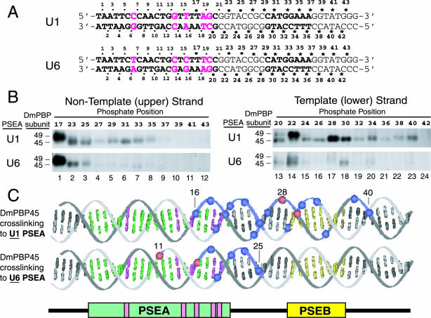
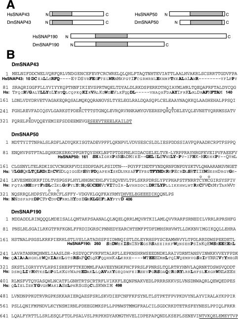
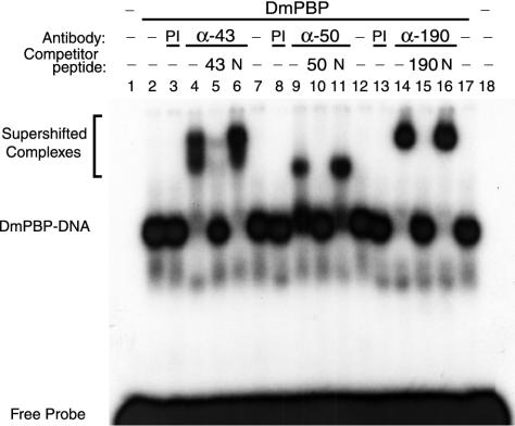
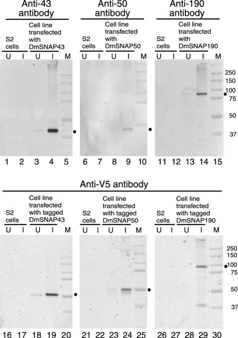
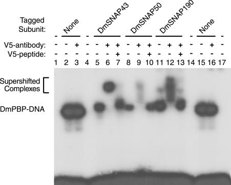
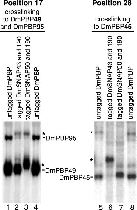
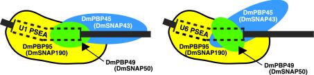
Transcription of snRNA genes by either RNA polymerase II (U1 to U5) or RNA polymerase III (U6) is dependent upon a proximal sequence element (PSE) located approximately 40 to 60 bp upstream of the transcription start site. In Drosophila melanogaster, RNA polymerase specificity is determined by as few as three nucleotide differences within the otherwise well-conserved 21-bp PSE. Previous photo-cross-linking studies revealed that the D. melanogaster PSE-binding protein, DmPBP, contains three subunits (DmPBP45, DmPBP49, and DmPBP95) that associate with the DNA to form complexes that are conformationally distinct depending upon whether the protein is bound to a U1 or a U6 PSE. We have identified and cloned the genes that code for these subunits of DmPBP by virtue of their similarity to three of the five subunits of SNAP(c), the human PBP. When expressed in S2 cells, each of the three cloned gene products is incorporated into a protein complex that functionally binds to a PSE. We also find that the conformational difference referred to above is particularly pronounced for DmPBP45, herein identified as the ortholog of human SNAP43. DmPBP45 cross-linked strongly to DNA for two turns of the DNA helix downstream of the U1 PSE, but it cross-linked strongly for only a half turn of the helix downstream of a U6 PSE. These substantial differences in the cross-linking pattern are consistent with those of a model in which conformational differences in DmPBP-DNA complexes lead to selective RNA polymerase recruitment to U1 and U6 promoters.


Figures

FIG. 1.
FIG. 1.
Site-specific protein-DNA photo-cross-linking of DmPBP to phosphate positions downstream of the PSEAs of D. melanogaster U1 and U6 genes. (A) Sequences of the relevant areas of the U1 and U6 photo-cross-linking probes. The sequences of the 21-bp U1 and U6 PSEAs are shown in boldface type. (Also shown in boldface type is the sequence of an 8-bp U1 PSEB, which is a second sequence fairly well conserved in D. melanogaster snRNA genes transcribed by RNA polymerase II [16, 38].) The five base pairs where the U1 and U6 PSEAs differ are indicated by magenta letters. Except for these five positions, the probes were identical in sequence to ensure that any differences in the cross-linking pattern were due solely to the five differences within the two PSEAs. The individual phosphate positions at which a cross-linker was incorporated are indicated by the dots, asterisks, and numbers above and below the sequences. The dots indicate positions for which data were reported in an earlier study (33). The asterisks indicate new positions not previously investigated; these 36 phosphate positions were individually derivatized to generate the 36 new probes used in this study. The larger dots indicate positions repeated in the present study as intensity standards and size markers to relate the new studies to the previous one. (B) Twenty-four different radiolabeled site-specific U1 probes and 24 different U6 probes were each incubated in separate reaction mixtures with DmPBP. Following UV irradiation and nuclease digestion, polypeptides that cross-linked to the DNA were detected by SDS-gel electrophoresis and autoradiography. Only the regions of the gels that correspond to the mobilities of DmPBP49 and DmPBP45 are shown. The left panels show the results of cross-linking to the nontemplate strand, and the right panels show cross-linking to the template strand. In each case, the upper panel illustrates the pattern for the U1 PSEA and the lower panel illustrates the pattern for the U6 PSEA. (C) Summary of DmPBP45 cross-linking to DNA probes that contain either the U1 PSEA or the U6 PSEA. Cross-linking data from Fig. 1B, together with previously reported data (33), are shown projected onto B-form DNA. Phosphate positions that cross-linked relatively strongly to the 45-kDa subunit of DmPBP are indicated by blue spheres, whereas the very strongest cross-linking positions are indicated by red spheres. Weak cross-links are not indicated. Base pairs that comprise the PSEA are shown in green and magenta, and those that comprise the U1 PSEB are shown in yellow. When DmPBP was bound to a U1 PSEA, phosphate positions between 16 and 40 cross-linked to DmPBP45, but when DmPBP was bound to a U6 PSEA, cross-linking to DmPBP45 was restricted to a region between phosphates 11 and 25. When the DNA is oriented as shown, the cross-links to DmPBP45 are mainly on the upper surface of the DNA helix.
FIG.2.
FIG.2.
The D. melanogaster genome codes for proteins with similarity to Homo sapiens SNAP43 (HsSNAP43), HsSNAP50, and HsSNAP190. (A) BLAST searches of the D. melanogaster nucleic acid database identified genes predicted to code for proteins with similarities to three of the human SNAP proteins. The rectangles indicate the relative lengths of the proteins from the N to the C termini, and the shaded areas indicate the regions of the homologous protein pairs that share greater than 26% identity and 42% similarity. (B) The amino acid sequences of the predicted DmSNAP43, DmSNAP50, and DmSNAP190 proteins are shown. Sequences from the homologous human SNAPs, in those regions where the corresponding proteins from the two species exhibit substantial similarity (shaded areas in panel A), are shown below the fly sequences. Dissimilar residues are indicated by small capital letters, chemically similar residues are indicated by large capital letters, and identical residues are indicated by boldface. The amino acid positions in the human SNAPs of the first and last residues shown are indicated by the numbers immediately before and after the human sequences (Hs). The underlined residues indicate the peptide sequences from the fly proteins that were used to generate antibodies. The sequence of the DmSNAP43 gene, cloned by us from both wild-type embryo DNA and S2 tissue culture cells, codes for a protein that differs at four amino acid positions from the sequence predicted in the D. melanogaster nucleic acid database. These four amino acids are indicated by asterisks above the DmSNAP43 sequence. The open circles above the DmSNAP50 sequence indicate four cysteines that may form a zinc finger and are conserved in homologous human, trypanosome, and roundworm proteins.
FIG. 3.
FIG. 3.
Antibodies against peptide sequences from the predicted DmSNAP proteins react with native DmPBP. Electrophoretic mobility shift assays were carried out by using DmPBP prepared from fruit fly embryos and a probe that contains U1 PSEA. Additional components were added as indicated above the individual lanes. Lanes 4 to 6, 9 to 11, and 14 to 16 contained antibodies from rabbits injected with synthetic peptides corresponding to sequences from the DmSNAP43 (α-43), DmSNAP50 (α-50), and DmSNAP190 (α-190) sequences, respectively (Fig. 2). Lanes 3, 8, and 13 contained preimmune sera (PI). Lanes 5, 10, and 15 contained excess synthetic peptide specific to the antibodies used in the same lane. Lanes 6, 11, and 16 contained nonspecific peptide (N).
FIG. 4.
FIG. 4.
Inducible overexpression of DmSNAP proteins in S2 cells. We generated S2 cell lines that were stably transfected with one of the DmSNAP genes under the control of the metallothionein promoter. Lysates from control S2 cells, or from stably transfected S2 cells, either without (lanes U) or with (lanes I) copper induction were analyzed by immunoblotting. Untagged DmSNAPs were detected by using specific antipeptide antibodies (upper panels) (Anti-43, Anti-50, and Anti-190 refer to antibodies to DmSNAP43, DmSNAP50, and DmSNAP190, respectively), whereas epitope-tagged versions of the proteins were analyzed by using anti-V5 antibodies (lower panels). Dots indicate the positions of bands that correspond to the gene products analyzed in each individual panel. Lanes M, molecular size markers.
FIG. 5.
FIG. 5.
Epitope-tagged DmSNAP proteins can be incorporated into DmPBP that functionally binds to DNA. Electrophoretic mobility shift assays were performed with DmPBP obtained from three different stably transfected S2 cell lines that each overexpressed all three DmSNAP subunits, but as indicated above the appropriate lanes (5 to 7, 8 to 10, and 11 to 13), only one of the subunits in each cell line possessed a V5 tag. Lanes 2, 3, 15, and 16 contained untagged DmPBP from fruit fly embryos. Anti-V5 antibody and V5 competitor peptide were added to reaction mixtures as indicated above the lanes.
FIG. 6.
FIG. 6.
Correlation of the DmSNAP gene products with the DmPBP subunits detected by photo-cross-linking. Photo-cross-linking was carried out with probes derivatized with a cross-linker at phosphate position 17 (probe reacts with DmPBP49 and DmPBP95 [lanes 1 to 4]) or at position 28 (probe reacts with DmPBP45 [lanes 5 to 8]). Protein components in the reaction mixtures were as follows: untagged DmPBP from embryos (lanes 1, 4, 5, and 8); protein from stably transfected S2 cell lines expressing V5/His6-tagged DmSNAP43, tagged DmSNAP190, and untagged DmSNAP50 (lanes 2 and 6); or protein from stably transfected S2 cell lines expressing V5/His6-tagged DmSNAP50, tagged DmSNAP190, and untagged DmSNAP43 (lanes 3 and 7). The asterisks indicate the positions of cross-linking products with reduced mobility due to the C-terminal polypeptide extension that contains the V5 and His6 epitopes. The dot indicates a nonspecific band observed only in the embryo protein preparation that is not competed by excess unlabeled probe (data not shown).
FIG. 7.
FIG. 7.
Schematic model of the subunits of DmPBP (DmSNAPc) bound in alternative conformations to D. melanogaster U1 and U6 PSEAs. The model is based upon past (5, 33) and present data. The three subunits of the PSEA-binding protein are shown in the approximate relative positions where they interact with the phosphate backbone of the DNA. In each case, the DNA is bent similarly toward the face of the DNA that interacts with DmPBP45 (DmSNAP43) (5). If this is considered to be the upper face of the PSEA, DmPBP49 (DmSNAP50) interacts primarily with the front face of the 3′ half of the PSEA, and DmPBP95 (DmSNAP190) interacts primarily with the front face of the 5′ half of the PSEA but with the bottom face of the 3′ half of the PSEA.

References

    1. Bai, L., Z. Wang, J.-B. Yoon, and R. G. Roeder. 1996. Cloning and characterization of the β subunit of human proximal sequence element-binding transcription factor and its involvement in transcription of small nuclear RNA genes by RNA polymerases II and III. Mol. Cell. Biol. 16:5419-5426. - PMC - PubMed
    1. Dahlberg, J. E., and E. Lund. 1988. The genes and transcription of the major small nuclear RNAs, p. 38-70. In M. L. Birnstiel (ed.), Structure and function of major and minor small nuclear ribonucleoprotein particles. Springer Verlag KG, Heidelberg, Federal Republic of Germany.
    1. Dahlberg, J. E., and E. Lund. 1991. How does III × II make U6? Science 254:1462-1463. - PubMed
    1. Das, A., and V. Bellofatto. 2003. RNA polymerase II-dependent transcription in trypanosomes is associated with a SNAP complex-like transcription factor. Proc. Natl. Acad. Sci. USA 100:80-85. - PMC - PubMed
    1. Hardin, S. B., C. J. Ortler, K. J. McNamara-Schroeder, and W. E. Stumph. 2000. Similarities and differences in the conformation of protein-DNA complexes at the U1 and U6 snRNA gene promoters. Nucleic Acids Res. 28:2771-2778. - PMC - PubMed

Publication types

MeSH terms