. 2004 Mar;24(5):1930-43.
doi: 10.1128/MCB.24.5.1930-1943.2004.

Replication protein A (RPA) phosphorylation prevents RPA association with replication centers

Affiliations

Replication protein A (RPA) phosphorylation prevents RPA association with replication centers

Vitaly M Vassin et al. Mol Cell Biol. 2004 Mar.

Abstract

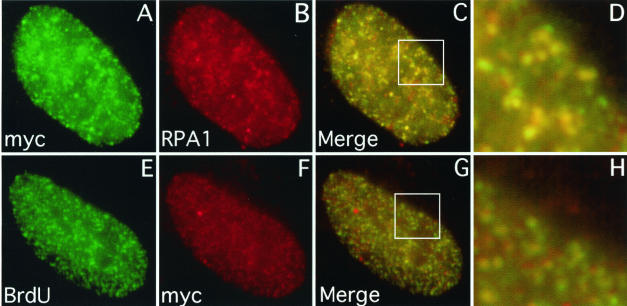
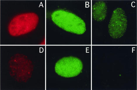
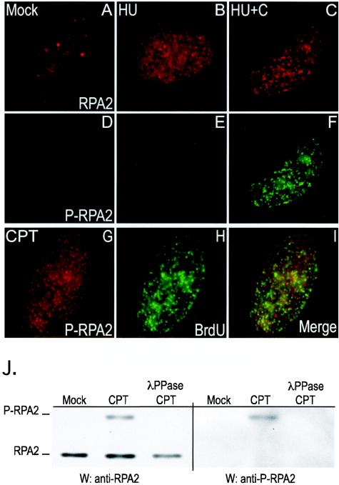
Mammalian replication protein A (RPA) undergoes DNA damage-dependent phosphorylation at numerous sites on the N terminus of the RPA2 subunit. To understand the functional significance of RPA phosphorylation, we expressed RPA2 variants in which the phosphorylation sites were converted to aspartate (RPA2(D)) or alanine (RPA2(A)). Although RPA2(D) was incorporated into RPA heterotrimers and supported simian virus 40 DNA replication in vitro, the RPA2(D) mutant was selectively unable to associate with replication centers in vivo. In cells containing greatly reduced levels of endogenous RPA2, RPA2(D) again did not localize to replication sites, indicating that the defect in supporting chromosomal DNA replication is not due to competition with the wild-type protein. Use of phosphospecific antibodies demonstrated that endogenous hyperphosphorylated RPA behaves similarly to RPA2(D). In contrast, under DNA damage or replication stress conditions, RPA2(D), like RPA2(A) and wild-type RPA2, was competent to associate with DNA damage foci as determined by colocalization with gamma-H2AX. We conclude that RPA2 phosphorylation prevents RPA association with replication centers in vivo and potentially serves as a marker for sites of DNA damage.


Figures

FIG. 1.
FIG. 1.
The myc-RPA2wt subunit colocalizes with endogenous RPA1 and DNA replication centers. U2-OS cells were transfected with a vector expressing myc-RPA2wt. To allow visualization of chromatin-bound myc-RPA, the cells were first extracted with 0.5% Triton X-100 for 5 min and then fixed with formaldehyde. (E to H) To detect sites of DNA replication, BrdU was added to the medium 10 min before the cells were prepared for epifluorescence microscopy. As indicated, the cells were then stained with anti-myc (A and F), anti-RPA1 (B), or anti-BrdU (E) antibody. The extent of myc-RPA2wt and endogenous RPA1 colocalization is shown (C), with enlargement of a particular nuclear region (boxed) (D). BrdU and myc-RPA2wt colocalization are similarly shown (G and H).
FIG. 2.
FIG. 2.
Lack of association of the RPA2D mutant with chromatin in unstressed cells. U2-OS cells were transfected with a vector expressing myc-RPA2wt (A to D), myc-RPA2D (E to H), or myc-RPA2A (I to L). (C, D, G, H, K, and L) To allow visualization of chromatin-bound myc-RPA, cells were first extracted with 0.5% Triton X-100 for 5 min and then fixed with formaldehyde (+ extraction). (A, B, E, F, I, and J) To assay for transfection efficiency, cells were also fixed without prior extraction (− extraction). The cells were then stained with anti-myc (B, D, F, H, J, and L) or anti-RPA1 (A, C, E, G, I, and K) antibody.
FIG. 3.
FIG. 3.
RPA2D-containing RPA heterotrimers are replication competent. (A) U2-OS cells were transfected with empty vector (lanes 1 and 4), myc-RPA2wt (lanes 2 and 5), or myc-RPA2D (lanes 3 and 6). Lysates were prepared from each batch of transfected cells, and the lysates were subjected to immunoprecipitation using anti-RPA1 antibodies. Immunoprecipitates (IP) (lanes 1 to 3) and aliquots of the lysates (lanes 4 to 6) were then analyzed for the presence of RPA2 by Western blotting analysis using RPA2 antibodies (which recognize both transfected and endogenous RPA2). (B and C) RPA heterotrimers that contained either RPA2wt (lane 1) or RPA2D (lane 2) were expressed in E. coli, purified, and analyzed by SDS-PAGE and Coomassie blue staining (B). The purified RPA was then assayed for the ability to support SV40 DNA replication in combination with an AS65 fraction purified from HeLa cells (51) (C). The open triangle shows that only background levels of DNA synthesis occur in reactions containing RPA2wt but lacking T antigen. Similar results were observed using RPA2D. (D) SV40 DNA replication reactions were performed in the presence of [α-32P]dCTP to label the replication products as described in Materials and Methods. The reaction mixtures contained 600 ng of either RPARPA2wt (lanes 1, 2, 4, and 5) or RPARPA2D (lanes 3 and 6) and either lacked T antigen (lanes 1 and 4) or contained 750 ng of T antigen (lanes 2, 3, 5, and 6). After isolation, the DNA replication products were first linearized by restriction digestion and then either mock treated (lanes 1 to 3) or incubated with DpnI to cleave nonreplicated DNA (lanes 4 to 6). The digestion products were then subjected to agarose gel electrophoresis, and the images of the ethidium bromide (EtBr)-stained gel (to show the total level of DNA) and the autoradiograph of the gel (to visualize 32P-labeled reaction products) are provided. The observed bands correspond to the linearized SV40 origin-containing plasmid.
FIG. 4.
FIG. 4.
Expression of RPA2D does not affect cell cycle progression. Cells transfected with myc-RPA2wt (A and B) or myc-RPA2D (C and D) were incubated with 10 μM BrdU for 30 min prior to harvest. The cells were then subjected to FACS analysis using a pairwise analysis of the levels of myc and BrdU signals. Transfected cells (with significant myc signals [boxed regions in panels A and C]) were further analyzed for the BrdU signal and the amount of DNA. (B and D) Fractions of cells in G1, S, and G2 phases. For each plot, the x and y axes indicate fluorescence intensities of the different signals.
FIG. 5.
FIG. 5.
Lack of RPA2D chromatin association in cells lacking endogenous RPA2. U2-OS cells were incubated with a control (i.e., scrambled) siRNA (A) or an siRNA specific for the 3′ noncoding region of the RPA2 mRNA (B to F). The cells were simultaneously cotransfected with an empty control vector (D), myc-RPA2wt (B and C), or myc-RPA2 D (E and F). Forty-eight hours posttransfection, the cells were extracted with 0.5% (vol/vol) Triton X-100 for 5 min prior to formaldehyde fixation to reveal RPA associated with chromatin (C and F) or were fixed to show total endogenous or transfected RPA2 (A, B, D, and E). The cells were then stained with anti-RPA2 (A and D) or anti-myc (panels B, C, E, and F) antibody and then visualized by epifluorescence microscopy. Cells with representative signals were chosen.
FIG. 6.
FIG. 6.
Increase in negative charge at the RPA2 N terminus decreases frequency of myc-positive cells. (A to F) Cells were transfected with myc-RPA2wt or myc-RPA2D41 as indicated. Representative epifluorescence images showing total myc-RPA2 staining (no extraction [A and B]), chromatin-bound myc-RPA2 (C and D), and chromatin-bound RPA1 (E and F) are shown. (G and H) Serines and threonine (boldface and underlined) at the potential phosphorylation sites in the N terminus of the RPA2 subunit were replaced with alanines (A) or aspartates (D) as indicated. Cells expressing these myc-tagged RPA2 mutants were then analyzed by immunofluorescence microscopy for the presence of myc-RPA2 bound to chromatin and, in parallel reactions, for transfection efficiency. The relative frequencies of myc-RPA2 positive cells were calculated as described in Materials and Methods.
FIG. 7.
FIG. 7.
RPA2D binds chromatin and colocalizes with RPA1 after CPT treatment. U2-OS cells were transfected with myc-RPA2wt (A to C), myc-RPA2D (D to F), or myc-RPA2A (G to I) vector. Forty-eight hours posttransfection, the cells were incubated with 1 μM CPT for 3 h, extracted with 0.5% (vol/vol) Triton X-100 for 5 min, fixed, and stained with anti-RPA1 (A, D, and G) and anti-myc (B, E, and H) antibodies. (C, F, and I) Colocalization of the two stains, enlarged from the boxed regions.
FIG. 8.
FIG. 8.
Collapse of DNA replication forks stimulates RPA loading to damaged DNA. (A to H) U2-OS cells were transfected with plasmids expressing either myc-RPA2wt or myc-RPA2D as indicated for 48 h. The cells were then treated with 2.5 mM HU for either 1 (A and E) or 3 (B and F) h in the absence of caffeine or treated with HU for 1 (C and G) or 3 (D and H) h in the presence of 5 mM caffeine. The cells were Triton X-100 extracted and fixed and then stained for myc-RPA. (I to L) Colocalization of myc-RPA2D with sites of DNA damage. U2-OS cells were transfected with a myc-RPA2D-expressing plasmid. Forty-eight hours posttransfection, the cells were treated with 2.5 mM HU for 3 h and then extracted with 0.5% Triton X-100 and fixed. The cells were stained with γ-H2AX (I) and myc-RPA (J). The staining pattern of a representative cell and the image of the merged staining patterns (K) are provided. (L) One section (boxed) of the composite image is shown enlarged to reveal the degree of signal overlap. (M) Graph showing the fractions of myc-RPA2-transfected cells with a significant chromatin-bound myc-RPA2wt (green bars) or myc-RPA2D (brown bars) signal. The fractions of cells showing chromatin-bound RPA were quantified by visual inspection of 100 to 200 cells. The bar graph values were calculated as described in Materials and Methods. Note that although RPA2wt staining is consistently detected in a greater fraction of cells than RPA2D staining, this result is expected because RPA2wt can be observed both at replication centers and at DNA damage foci while RPA2D localizes only to damage foci. (N) Effects of stress and caffeine treatment on endogenous RPA2 phosphorylation. U2-OS cells were mock treated (mock) or treated with either 1 μM CPT for 1 or 3 h, 2.5 mM HU for 1 or 3 h, 5 mM caffeine (c) for 3 h, 2.5 mM HU for 1 or 3 h in the presence of 5 mM caffeine or with 7 μM aphidicolin (A) for 3 h and 7 μM aphidicolin for 1 or 3 h in the presence of 5 mM caffeine. The band labeled RPA2′ represents nonphosphorylated RPA, while P-RPA2 indicates the position of phosphorylated RPA.
FIG. 9.
FIG. 9.
Endogenous phosphorylated RPA (P-RPA2) does not localize to sites of DNA synthesis. (A to F) U2-OS cells were either mock treated (A and D) or treated with 2.5 mM HU for 3 h (B and E) or with 2.5 mM HU and 5 mM caffeine for 3 h (C and F). The cells were extracted to visualize the chromatin-bound form of RPA, fixed, and then stained either with anti-RPA2 (A to C) or anti-pSer4/pSer8-RPA2 (D to F) antibody. (G to I) U2-OS cells were either mock treated or treated with 1 μM CPT for 30 min, followed by an additional 2.5-h incubation in medium lacking CPT. The cells were incubated with 10 μM BrdU for 15 min prior to being processed. The cells were then extracted to visualize the chromatin-bound form of RPA, fixed, and stained either with anti-pSer4/pSer8-RPA2 (G) or anti-BrdU (H) antibody. (I) Merged staining pattern. (J) Extracts prepared from mock-treated or CPT-treated (1 μM for 3 h) cells were subjected to Western blotting (W) analyses using either anti-RPA2 (34A) monoclonal antibody (anti-RPA2) or a rabbit anti-pSer4/pSer8-RPA2 antibody (anti-P-RPA2). CPT-treated extracts were also incubated with λ protein phosphatase (λPPase), as indicated.

Similar articles

Cited by

References

    1. Abraham, R. T. 2001. Cell cycle checkpoint signaling through the ATM and ATR kinases. Genes Dev. 15:2177-2196. - PubMed
    1. Ariza, R. R., S. M. Keyse, J. G. Moggs, and R. D. Wood. 1996. Reversible protein phosphorylation modulates nucleotide excision repair of damaged DNA by human cell extracts. Nucleic Acids Res. 24:433-440. - PMC - PubMed
    1. Bartek, J., and J. Lukas. 2001. Mammalian G1- and S-phase checkpoints in response to DNA damage. Curr. Opin. Cell Biol. 13:738-747. - PubMed
    1. Binz, S. K., Y. Lao, D. F. Lowry, and M. S. Wold. 2003. The phosphorylation domain of the 32-kDa subunit of replication protein A modulates RPA-DNA interactions: evidence for an intersubunit interaction. J. Biol. Chem. 278:35584-35591. - PubMed
    1. Borowiec, J. 1992. Inhibition of structural changes in the simian virus 40 core origin of replication by mutation of essential origin sequences. J. Virol. 66:5248-5255. - PMC - PubMed

Publication types

LinkOut - more resources