. 2004 Mar;24(5):1956-67.
doi: 10.1128/MCB.24.5.1956-1967.2004.

Barrier proteins remodel and modify chromatin to restrict silenced domains

Affiliations

Barrier proteins remodel and modify chromatin to restrict silenced domains

Masaya Oki et al. Mol Cell Biol. 2004 Mar.

Abstract

Transcriptionally active and inactive domains are frequently found adjacent to one another in the eukaryotic nucleus. To better understand the underlying mechanisms by which domains maintain opposing transcription patterns, we performed a systematic genomewide screen for proteins that may block the spread of silencing in yeast. This analysis identified numerous proteins with efficient silencing blocking activities, and some of these have previously been shown to be involved in chromatin dynamics. We isolated subunits of Swi/Snf, mediator, and TFIID, as well as subunits of the Sas-I, SAGA, NuA3, NuA4, Spt10p, Rad6p, and Dot1p complexes, as barrier proteins. We demonstrate that histone acetylation and chromatin remodeling occurred at the barrier and correlated with a block to the spread of silencing. Our data suggest that multiple overlapping mechanisms were involved in delimiting silenced and active domains in vivo.


Figures
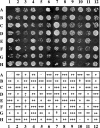
FIG. 1.
FIG. 1.
Systematic screening with GBD-fused library. ROY1864 (MATα hmrΔ::bgl-bcl) containing pRO486 (HMR-EΔI plus GAL4 bs) was individually transformed with a GBD-fused ORF library and grown in selective liquid culture in microtiter plates. Cells were transferred to YMD plates with mating lawns to assay for expression of the MATa1 gene at HMR. The growth of diploid colonies is shown. We classified each clone into five categories. −, 0 diploid colonies (very strong barrier); ±, 1 to 9 colonies (strong barrier); +, 10 to 20 (moderate barrier); ++, >21 diploid colonies (weak barrier); +++, no barrier.
FIG. 2.
FIG. 2.
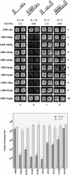
(Top) Relative barrier activity of the GBD chimeras. We used patch mating assays to analyze the ability of clones to block silencing but not disrupt silencing. ROY1864 was transformed with any of four different URA3-containing reporter plasmids: pRO363 (HMR-EΔI), pRO486 (HMR-EΔI plus GAL4 bs), pRO4 (HMR-E+I), and pRO651 (HMR-E+I plus GAL4 bs). Each of these strains was cotransformed with a second TRP1-containing plasmid containing variousGBD fusion chimeras as indicated. Cells were grown on selective medium (YMD lacking both Trp and Ura) and were analyzed by patch mating against tester lawns (JRY19a) for silencing of the MATa1 gene. Growth of diploid cells indicates silencing of the reporter gene, while absence of any growth indicates activation of the reporter gene due to blocking of silencing. The chimera plasmids used are GBD alone (pGBK-RC), GBD-Snf6p (pRO586), GBD-Taf47p (pRO587), GBD-Ada1p (pRO588), GBD-Ada2p (pRO592), GBD-Sas2p (pRO590), GBD-Sas5p (pRO591), GBD-Clb1p (pRO594), GBD-Dot1p (pRO637),and GBD-Nup2p (pRO635). (Bottom) Quantitative mating analyses of various GBD chimeras in strain ROY1864 containing either pRO486 (E only) or pRO651 (E & I) were performed as described previously (12, 39). The data are presented as diploid CFU and are mean values from three independent experiments carried out in parallel.
FIG. 3.
FIG. 3.
Boundary activity in disruption strains. We used patch mating assays to analyze the ability of GBD barrier chimeras to block silencing in strains from which specific genes were deleted. Each strain was transformed with the different plasmids as described in the legend of Fig. 2 and assayed on the same plate. (−) and (+), constructs without and with Aal4p binding sites, respectively; wt, wild type.
FIG. 4.
FIG. 4.
(Top) Barrier activity at telomere VIIL. Strains YDS631 (adh4::URA3-telVII-L) and YDS634 (adh4::URA3-4xGAL4 bs-telVII-L) (9) were transformed with GBD fusion chimera plasmids as described in the legend of Fig. 2. Cells were grown on selective liquid media (YMD lacking Trp) and spotted as 10-fold serial dilutions on YMD plates lacking uracil and tryptophan or containing 5-FOA as described previously (28). (Bottom) Strain KIY54 (25) and ROY 2770 were transformed with the GBD fusion chimera plasmids described in the legend of Fig. 2A. The strains were grown in liquid YMD-HC medium with selection (−trp) overnight, and the cells were spotted as 10-fold serial dilutions on YMD-HC plates lacking adenine and tryptophan or containing 5 μg of adenine per ml but lacking tryptophan as described previously (28). The plates were photographed after 2 days.
FIG. 4.
FIG. 4.
(Top) Barrier activity at telomere VIIL. Strains YDS631 (adh4::URA3-telVII-L) and YDS634 (adh4::URA3-4xGAL4 bs-telVII-L) (9) were transformed with GBD fusion chimera plasmids as described in the legend of Fig. 2. Cells were grown on selective liquid media (YMD lacking Trp) and spotted as 10-fold serial dilutions on YMD plates lacking uracil and tryptophan or containing 5-FOA as described previously (28). (Bottom) Strain KIY54 (25) and ROY 2770 were transformed with the GBD fusion chimera plasmids described in the legend of Fig. 2A. The strains were grown in liquid YMD-HC medium with selection (−trp) overnight, and the cells were spotted as 10-fold serial dilutions on YMD-HC plates lacking adenine and tryptophan or containing 5 μg of adenine per ml but lacking tryptophan as described previously (28). The plates were photographed after 2 days.
FIG. 5.
FIG. 5.
Analysis of chromatin structure around the barrier. Strain ROY2042 containing HMRΔI with four Gal4p binding sites located between HMR-E and the MATa1 gene was transformed with pGBK-RC (GBD alone), pRO586 (GBD-Snf6p), or pRO590 (GBD-Sas2p). Nuclei were prepared from these strains and digested for various lengths of time with micrococcal nuclease. The deproteinized DNA was then digested with BglII, and the DNA was resolved on an agarose gel, blotted onto membranes, and probed with specific probes. This figure shows five time points of digestion by micrococcal nuclease for each strain. The region between HMR-E and the start of the MATa1 gene is shown on the right with a densitometric scan of the digestion profile.
FIG. 6.
FIG. 6.
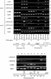
(Top) Histone H4 acetylation analysis around the Gal4p binding site barrier. Chromatin immunoprecipitation analysis was performed with antibodies against Sir3p, acetyl-histone H4, acetyl-H4K16, or acetyl-H3 K14. Strains ROY2042 (SAS2) and ROY2243 (sas2Δ) containing HMRΔI with four Gal4p binding sites located between HMR-E and the MATa1 gene were transformed with pGBK-RC (GBD only), pRO586 (GBD-Snf6p), or pRO590 (GBD-Sas2p). All six strains were grown in liquid culture prior to cross-linking and immunoprecipitations. PCR probe A analyzed a region between HMR-E and the barrier, probe B was located between the barrier and the MATa1 gene, and probe C was located in the coding region of the MATa1 gene. Probe D analyzed a region 7.5 kb from telomere 6R, and probe E analyzed a region 500 bp from telomere 6R. (Bottom) Strain JRY4013 was grown in liquid culture prior to cross-linking. Immunoprecipitations were performed with antibodies against Sir3p, acetyl-H4K16, and acetyl-H3K14. The probes used were as described previously (73).

Similar articles

Cited by

References

    1. Abraham, J., K. A. Nasmyth, J. N. Strathern, A. J. Klar, and J. B. Hicks. 1984. Regulation of mating-type information in yeast. Negative control requiring sequences both 5′ and 3′ to the regulated region. J. Mol. Biol. 176:307-331. - PubMed
    1. Aparicio, O. M., and D. E. Gottschling. 1994. Overcoming telomeric silencing: a trans-activator competes to establish gene expression in a cell cycle-dependent way. Genes Dev. 8:1133-1146. - PubMed
    1. Bi, X. 2002. Domains of gene silencing near the left end of chromosome III in Saccharomyces cerevisiae. Genetics 160:1401-1407. - PMC - PubMed
    1. Bi, X., M. Braunstein, G. J. Shei, and J. R. Broach. 1999. The yeast HML I silencer defines a heterochromatin domain boundary by directional establishment of silencing. Proc. Natl. Acad. Sci. USA 96:11934-11939. - PMC - PubMed
    1. Bi, X., and J. R. Broach. 1997. DNA in transcriptionally silent chromatin assumes a distinct topology that is sensitive to cell cycle progression. Mol. Cell. Biol. 17:7077-7087. - PMC - PubMed

Publication types

MeSH terms