. 2004 Mar;24(5):1990-9.
doi: 10.1128/MCB.24.5.1990-1999.2004.

Persistent signaling by dysregulated thrombin receptor trafficking promotes breast carcinoma cell invasion

Affiliations

Persistent signaling by dysregulated thrombin receptor trafficking promotes breast carcinoma cell invasion

Michelle A Booden et al. Mol Cell Biol. 2004 Mar.

Abstract

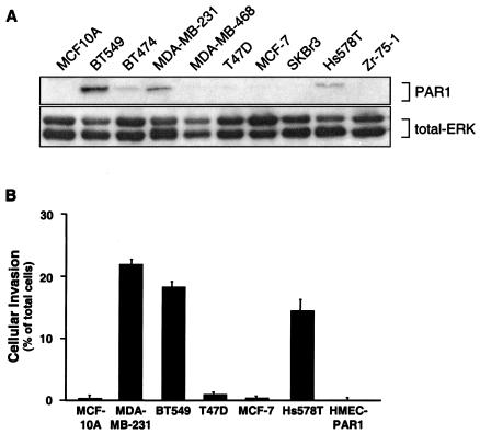
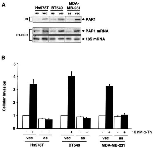
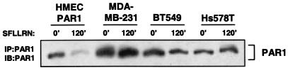
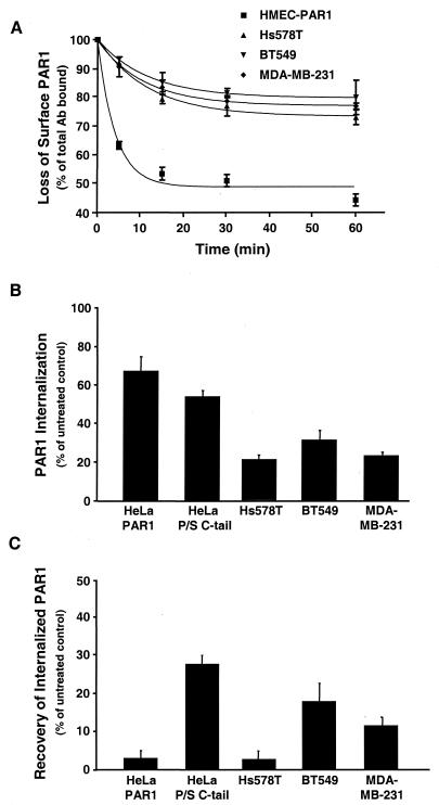
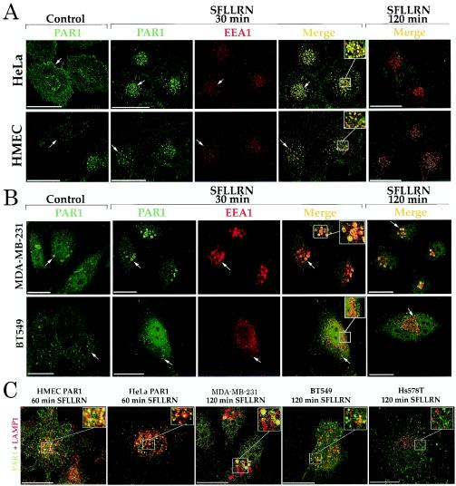
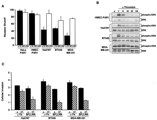
Increased expression of protease-activated receptor 1 (PAR1), a G protein-coupled receptor for thrombin, has previously been correlated with breast carcinoma cell invasion. PAR1 is irreversibly proteolytically activated, internalized, and sorted directly to lysosomes, a critical process for the termination of signaling. We determined that activated PAR1 trafficking is severely altered in metastatic breast carcinoma cells but not in nonmetastatic or normal breast epithelial cells. Consequently, the proteolytically activated receptor is not sorted to lysosomes and degraded. Altered trafficking of proteolytically activated PAR1 caused sustained activation of phosphoinositide hydrolysis and extracellular signal-regulated kinase signaling, even after thrombin withdrawal, and enhanced cellular invasion. Thus, our results reveal that a novel alteration in trafficking of activated PAR1 causes persistent signaling and, in addition to other processes and proteins, contributes to breast carcinoma cell invasion.


Figures

FIG. 1.
FIG. 1.
PAR1 protein is overexpressed in invasive breast carcinoma cell lines. (A) Lysates prepared from multiple breast carcinoma cell lines were resolved by SDS-15% PAGE (50 μg of total protein), transferred, and immunoblotted with an anti-PAR1 monoclonal antibody. Immunoblotting these same membranes for total ERK with a polyclonal anti-p42/44 ERK antibody served as a control for equal loading. (B) Basal invasion activity of breast carcinoma cell lines was measured using a Matrigel invasion assay. All of the cell lines were serum starved for a minimum of 24 h, and cellular invasion was assessed by the addition of α-Th to the upper well only to stimulate PAR1 activation. The data are expressed as the percentage of cells that invaded compared to the total number of cells seeded in the upper chamber and are reported as the mean percentage plus the standard error of the mean of at least two independent experiments for MCF10A, HMEC-PAR1, and T47D cells; three independent experiments for MCF-7 and SKBR3 cells; and six independent experiments for MDA-MB-231, Hs578T, and BT549. The average numbers of cells invading under basal conditions in a given experiment were between 0 and 100 cells for MCF-10A, T47D, MCF-7, HMEC-PAR1 and SKBr3 and between 200 and 600 cells for MDA-MB-231, BT549, and Hs578T.
FIG. 2.
FIG. 2.
Expression and activation of PAR1 enhances thrombin-mediated breast carcinoma cell invasion. (A) Lysates prepared from cells stably expressing PAR1 antisense (as) cDNA or empty vector (vec) were resolved by SDS-PAGE (50 μg of total protein) and immunoblotted using a monoclonal PAR1 antibody. RT-PCR was used to assess PAR1 mRNA levels in antisense and vector control-expressing cells, and 18S mRNA was amplified as a control. (B) MDA-MB-231, BT549, and Hs578T cells expressing empty vector or PAR1 antisense cDNA were serum starved for a minimum of 24 h and then incubated in the absence or presence of 10 nM α-Th, and cellular invasion was assessed. The data are expressed as the increase (n-fold) in cells invaded compared to untreated controls and represent the means plus standard errors of the mean of at least three independent experiments. The average numbers of cells invading after stimulation with 10 nM α-Th in a given experiment were between 0 and 20 for HMEC-PAR1 and between 600 and 1,100 for MDA-MB-231, BT549, and Hs578T.
FIG. 3.
FIG. 3.
Defective degradation of activated PAR1 in invasive breast carcinoma cell lines. Cells plated at a density of 106 were incubated in the absence (0′) or presence (120′) of 50 μM SFLLRN for 120 min at 37°C. The cells were lysed and immunoprecipitated (IP) with monoclonal anti-PAR1 antibody. The immunoprecipitates were resolved by SDS-9% PAGE, transferred, and immunoblotted (IB) with an anti-PAR1 polyclonal antibody.
FIG.4.
FIG.4.
Agonist-induced internalization and recycling of PAR1 are perturbed in invasive breast carcinoma cells. (A) Cells were incubated in the absence or presence of 10 nM α-Th for the indicated times at 37°C and fixed, and the amount of PAR1 remaining on the cell surface was measured by enzyme-linked immunosorbent assay. The results are expressed as the mean percentage (± standard error of the mean [SEM]) of total antibody bound to untreated controls in two separate experiments in which duplicate determinations were made. (B and C) Cells labeled with anti-PAR1 antibody were incubated in the absence or presence of 50 μM SFLLRN for 60 min. The cells were then trypsin treated, and the amount of receptor-bound antibody reappearing at the cell surface was measured after 30 min. The amount of antibody remaining on the cell surface following trypsin treatment was between 2 and 8% of total binding and was subtracted to obtain the values shown. The results are the amounts of surface-bound antibody detected after agonist incubation at 37°C (B) and after 30 min of recovery (C) and are expressed as the mean percentages (±SEM) of total antibody bound to untreated control cells.
FIG. 5.
FIG. 5.
PAR1 trafficking is dysregulated in invasive breast carcinoma cells. (A) HeLa- and HMEC-PAR1 cells were preincubated with anti-PAR1 antibody at 4°C, washed, and then incubated in the absence (Control) or presence of 50 μM SFLLRN for 30 or 120 min at 37°C. The cells were fixed, permeabilized, and immunostained for PAR1 (green) and EEA1 (red). (B) MDA-MB-231 and BT549 cells were treated as described for panel A. (C) Cells labeled with anti-PAR1 antibody were agonist treated for either 60 or 120 min and then coimmunostained for PAR1 (green) and lysosomal marker protein LAMP1 (red). The images are representative of >100 cells examined in at least three independent experiments. The insets are magnifications of the boxed areas. Scale bar, 19.4 μm. Arrows indicate staining of PAR1-positive intracellular vesicles.
FIG. 6.
FIG. 6.
Persistent signaling by activated PAR1 promotes invasion by breast carcinoma cell lines. (A) Cells labeled with [myo-3H]inositol were incubated with 50 μM SFLLRN or 10 nM α-Th for 60 min at 37°C in the absence of LiCl. Agonists were removed, and the cells were washed with DMEM containing 0.5 U of hirudin/ml and incubated with DMEM (20 mM LiCl and hirudin) at 37°C for an additional 60 min, at which time accumulated 3H-labeled IPs were measured. The results shown are normalized to the amount of IP species generated when agonist was added with LiCl and are expressed as the mean percent (plus standard error of the mean [SEM]) shutoff of at least two separate experiments. (B) Cells were serum starved for 48 to 72 h and incubated in the presence or absence of 10 nM α-Th for the indicated times (in minutes) at 37°C. Cell lysates were prepared, resolved by SDS-PAGE, and immunoblotted with an anti-phospho-p42/44 ERK antibody. The same membranes were immunoblotted with anti-p42/44 antibody to control for total loading. (C) Cells added to the upper chamber were seeded in medium containing 0.1% BSA and either 10 nM α-Th or 50 μM SFLLRN, and the number of invading cells was determined (+) as described in the legends to Fig. 1 and 2. Other cells were pretreated with α-Th or SFLLRN for 60 min at 37°C, agonists were removed, and the cells were washed with DMEM containing 0.5 U of hirudin/ml and then seeded in the upper chamber in medium containing only 0.1% BSA, and the number of invaded cells was determined (+/−). The data are expressed as the increase (n-fold; mean + SEM) in cellular invasion induced by agonists compared to untreated control cells in at least three separate experiments.

References

    1. Albini, A., Y. Iwamoto, H. K. Kleinman, G. R. Martin, S. A. Aaronson, J. M. Kozlowski, and R. N. McEwan. 1987. A rapid in vitro assay for quantitating the invasive potential of tumor cells. Cancer Res. 47:3239-3245. - PubMed
    1. Bar-Sagi, D., and J. R. Feramisco. 1986. Induction of membrane ruffling and fluid-phase pinocytosis in quiescent fibroblasts by ras proteins. Science 233:1061-1068. - PubMed
    1. Bissell, M. J., and D. Radisky. 2001. Putting tumours in context. Nat. Rev. Cancer 1:46-54. - PMC - PubMed
    1. Christoforidis, S., H. M. McBride, R. D. Burgoyne, and M. Zerial. 1999. The Rab5 effector EEA1 is a core component of endosome docking. Nature 397:621-625. - PubMed
    1. Clark, G. J., and C. J. Der. 1995. Aberrant function of the Ras signal transduction pathway in human breast cancer. Breast Cancer Res. Treat. 35:133-144. - PubMed

Publication types