

Id2 is dispensable for Myc-induced epidermal neoplasia
- PMID: 14966287
- PMCID: PMC350569
- DOI: 10.1128/MCB.24.5.2083-2090.2004
Id2 is dispensable for Myc-induced epidermal neoplasia
Abstract
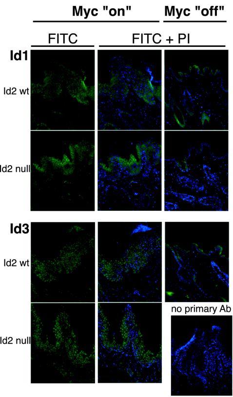
We have previously described a transgenic mouse model of epidermal neoplasia wherein expression of a switchable form of c-Myc, MycER(TAM), is targeted to the postmitotic suprabasal keratinocytes of murine epidermis via the involucrin promoter. Sustained activation of c-MycER(TAM) results in a progressive neoplastic phenotype characterized by aberrant ectopic proliferation and delayed differentiation of suprabasal keratinocytes, culminating in papillomatosis. Transcription of the Id2 gene is regulated by Myc family proteins. Moreover, Id2 is implicated as a pivotal determinant of cell fate in multiple lineages and has a demonstrated role in mediating Myc-dependent cell proliferation in vitro through its interaction with retinoblastoma protein. Using Id2 nullizygous mice, we assessed in vivo the requirement for Id2 in mediating Myc-induced papilloma formation in skin. We show that absence of Id2 has no discernible impact on any measurable attribute of Myc function or on the timing or extent of eventual tumor formation. Thus, our data argue against any essential role for Id2 in mediating Myc action in vivo.
Figures


Similar articles
-
Reversible activation of c-Myc in skin: induction of a complex neoplastic phenotype by a single oncogenic lesion.Mol Cell. 1999 May;3(5):565-77. doi: 10.1016/s1097-2765(00)80350-0. Mol Cell. 1999. PMID: 10360173
-
Omomyc expression in skin prevents Myc-induced papillomatosis.Cell Death Differ. 2004 Sep;11(9):1038-45. doi: 10.1038/sj.cdd.4401443. Cell Death Differ. 2004. PMID: 15143346
-
Id2 is a retinoblastoma protein target and mediates signalling by Myc oncoproteins.Nature. 2000 Oct 5;407(6804):592-8. doi: 10.1038/35036504. Nature. 2000. PMID: 11034201
-
MYC in mammalian epidermis: how can an oncogene stimulate differentiation?Nat Rev Cancer. 2008 Mar;8(3):234-42. doi: 10.1038/nrc2328. Nat Rev Cancer. 2008. PMID: 18292777 Free PMC article. Review.
-
The Max transcription factor network: involvement of Mad in differentiation and an approach to identification of target genes.Cold Spring Harb Symp Quant Biol. 1994;59:109-16. doi: 10.1101/sqb.1994.059.01.014. Cold Spring Harb Symp Quant Biol. 1994. PMID: 7587059 Review.
Cited by
-
The Id-protein family in developmental and cancer-associated pathways.Cell Commun Signal. 2017 Jan 25;15(1):7. doi: 10.1186/s12964-016-0161-y. Cell Commun Signal. 2017. PMID: 28122577 Free PMC article. Review.
-
siRNA directed against c-Myc inhibits proliferation and downregulates human telomerase reverse transcriptase in human colon cancer Colo 320 cells.J Exp Clin Cancer Res. 2008 Aug 12;27(1):27. doi: 10.1186/1756-9966-27-27. J Exp Clin Cancer Res. 2008. Retraction in: J Exp Clin Cancer Res. 2009 Jul 16;28:101. doi: 10.1186/1756-9966-28-101. PMID: 18694522 Free PMC article. Retracted.
-
The Smad7-Skp2 complex orchestrates Myc stability, impacting on the cytostatic effect of TGF-β.J Cell Sci. 2014 Jan 15;127(Pt 2):411-21. doi: 10.1242/jcs.136028. Epub 2013 Nov 20. J Cell Sci. 2014. PMID: 24259667 Free PMC article.
-
Genetic analysis of myc and telomerase interactions in vivo.Mol Cell Biol. 2006 Aug;26(16):6130-8. doi: 10.1128/MCB.00543-06. Mol Cell Biol. 2006. PMID: 16880523 Free PMC article.
-
Sequestration of E12/E47 and suppression of p27KIP1 play a role in Id2-induced proliferation and tumorigenesis.Carcinogenesis. 2009 Jul;30(7):1252-9. doi: 10.1093/carcin/bgp115. Epub 2009 May 18. Carcinogenesis. 2009. PMID: 19451188 Free PMC article.
References
-
- Benezra, R. 2001. Role of Id proteins in embryonic and tumor angiogenesis. Trends Cardiovasc. Med. 11:237-241. - PubMed
-
- Benezra, R., R. L. Davis, D. Lockshon, D. L. Turner, and H. Weintraub. 1990. The protein Id: a negative regulator of helix-loop-helix DNA binding proteins. Cell 61:49-59. - PubMed
-
- Benezra, R., S. Rafii, and D. Lyden. 2001. The Id proteins and angiogenesis. Oncogene 20:8334-8341. - PubMed
-
- Brysk, M. M., I. Arany, H. Brysk, S. H. Chen, K. H. Calhoun, and S. K. Tyring. 1995. Gene expression of markers associated with proliferation and differentiation in human keratinocytes cultured from epidermis and from buccal mucosa. Arch. Oral Biol. 40:855-862. - PubMed
Publication types
MeSH terms
Substances
Grants and funding
LinkOut - more resources
Full Text Sources
Other Literature Sources
Medical
Molecular Biology Databases