. 2004 Mar;24(5):2181-9.
doi: 10.1128/MCB.24.5.2181-2189.2004.

Control of cyclin G2 mRNA expression by forkhead transcription factors: novel mechanism for cell cycle control by phosphoinositide 3-kinase and forkhead

Affiliations

Control of cyclin G2 mRNA expression by forkhead transcription factors: novel mechanism for cell cycle control by phosphoinositide 3-kinase and forkhead

Lorena Martínez-Gac et al. Mol Cell Biol. 2004 Mar.

Abstract

Cyclin G2 is an unconventional cyclin highly expressed in postmitotic cells. Unlike classical cyclins that promote cell cycle progression, cyclin G2 blocks cell cycle entry. Here we studied the mechanisms that regulate cyclin G2 mRNA expression during the cell cycle. Analysis of synchronized NIH 3T3 cell cultures showed elevated cyclin G2 mRNA expression levels at G(0), with a considerable reduction as cells enter cell cycle. Downregulation of cyclin G2 mRNA levels requires activation of phosphoinositide 3-kinase, suggesting that this enzyme controls cyclin G2 mRNA expression. Because the phosphoinositide 3-kinase pathway inhibits the FoxO family of forkhead transcription factors, we examined the involvement of these factors in the regulation of cyclin G2 expression. We show that active forms of the forkhead transcription factor FoxO3a (FKHRL1) increase cyclin G2 mRNA levels. Cyclin G2 has forkhead consensus motifs in its promoter, which are transactivated by constitutive active FoxO3a forms. Finally, interference with forkhead-mediated transcription by overexpression of an inactive form decreases cyclin G2 mRNA expression levels. These results show that FoxO genes regulate cyclin G2 expression, illustrating a new role for phosphoinositide 3-kinase and FoxO transcription factors in the control of cell cycle entry.


Figures
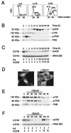
FIG. 1.
FIG. 1.
Cyclin G2 mRNA levels are maximal in G0. (A to C) NIH 3T3 cells were arrested by confluence and then released by low-density replating in medium with serum for different time periods. (A) Representative cell cycle profiles of cells 0, 18, and 23 h after release, showing the percentage of cells in G0/G1, S, and G2/M. (B) Western blot analysis of the NIH 3T3 cells entering the cell cycle synchronously, using anti-cyclin D3 (cycD3), anti-p130, anti-phospho Ser 473-PKB (pPKB), or anti-PKB antibody. (C) Northern blot analysis of cyclin G2 expression. Total RNA was extracted from NIH 3T3 cells (same as in panels A and B), resolved in agarose gels (10 μg), transferred, and hybridized with a probe for cyclin G2 or 28S rRNA. The percentage of cells in the S and G2/M phases is indicated. (D) Representative FKHRL1 (FoxO3a) immunofluorescence staining of NIH 3T3 cells deprived of serum for 18 h (−serum) or deprived of serum for 18 h and then incubated with serum for 1 h (+serum). (E and F) NIH 3T3 cells deprived of serum for 18 h (time 0), deprived of serum for 18 h and then incubated with serum for different times (indicated in minutes), or arrested in the G2 or M phase. (E) Western blot analysis using the indicated antibodies. (F) Northern blot analysis of cyclin G2 expression. Total RNA was extracted from aliquots of the cells used in panel E. The percentage of cells in the S and G2/M phases is indicated. The figure illustrates a representative experiment of at least three with similar results.
FIG. 2.
FIG. 2.
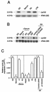
Downregulation of cyclin G2 mRNA levels in G1 requires PI3K activation. (A and B) Northern blot analysis for cyclin G2 expression in total RNA from NIH 3T3 cells incubated with different stimuli. (A) Cells were G0 arrested and stimulated (3 h) with 10% serum, 10-ng/ml LPA, or 100 μM PA. (B) Cells were G0 arrested and stimulated (3 h) with 15-ng/ml PDGF or 20-ng/ml IGF. Other samples were preincubated (1 h) with 10 μM LY294002 (LY) or 2 μM PD98059 (PD) and subsequently treated with 10% serum (3 h). Other samples were transfected with control cDNA or a vector encoding p110-CAAX (indicated); after incubation (8 h) in medium with serum, cells were cultured in the absence of serum for 18 h (G0) prior to analysis. (C) Q-PCR analysis of cyclin G2 expression. cDNA was obtained from cells treated as in panels A and B. For each condition, the amount of cyclin G2 cDNA relative to that of actin was estimated according to the Ct, which defines the PCR cycle number at which the signal reaches a defined value. The relative amount of cyclin G2 was calculated according to the formula ΔCtcyclin G2 = Ctcyclin G2 − Ctactin. Because maximal cyclin G2 expression is found in G0, relative expression under the different conditions (x) was compared to that of G0 by using the formula ΔΔCt = ΔCtcyclin G2 (G0) − ΔCtcyclin G2 (x). The mean ± standard deviation of three experiments is shown.
FIG. 3.
FIG. 3.
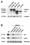
Activation of FoxO genes induces cyclin G2 mRNA expression. (A and B) NIH 3T3 cells were transfected with cDNA encoding FoxO3a wt (HA-FKHRL1) or active forms (HA-FKHRL1A3) or FoxO4 wt (HA-AFX) or active forms (HA-AFXA3). (A) Expression of FoxO TFs was examined by Western blotting with an anti-HA antibody. α-Tubulin expression was used as a control for equivalent loading. (B) After transfection, a fraction of the cells (same as in panel A) were incubated (8 h) in medium with serum, starved for 18 h in serum-free medium, and then examined (G0) or incubated (3 h) with 10% serum prior to analysis. Cell lysates were analyzed by Northern blotting with probes specific for p27kip, cyclin G2, or 28S rRNA.
FIG. 4.
FIG. 4.
Interfering mutants of FoxO genes downregulate cyclin G2 mRNA expression in G0. NIH 3T3 cells were transiently transfected with a control vector or with HA-ΔDBAFX. (A) Cells were cultured for 24 h. HA-ΔDBAFX expression was examined by Western blotting with an anti-HA antibody. α-Tubulin expression was analyzed as a loading control. (B) After transfection, a fraction of the cells (same as in panel A) were incubated (8 h) in medium with serum and then were cultured in the absence of serum for 18 h (G0) and then collected and examined. Untransfected control cells were cultured in the absence of serum (18 h) and then incubated with serum (3 h) or preincubated with LY294002 (1 h) prior to serum treatment (3 h). Total RNA was examined by Northern blot analysis for cyclin G2 mRNA expression relative to 28S rRNA levels. A representative experiment is shown of three experiments performed with similar results.
FIG. 5.
FIG. 5.
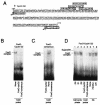
FoxO transcription factors bind to the cyclin G2 promoter. (A) Murine cyclin G2 promoter region containing the following FoxO TF consensus motifs: FKH1 consensus RYAAACAWW, Xenopus FKH consensus WRARYMAATA, IGFBP1 FKH-responsive promoter element AAAACAAACT (R = A/G, Y = T/C, W = A/T, M = A/C, K = T/G). Black dots indicate mismatches. Numbers indicate the position in the promoter relative to the transcription initiation site. (B and C) Electrophoretic mobility shift assays were performed with DG75 nuclear extracts and radiolabeled oligonucleotides representing the clustered FoxO consensus binding sites in the cyclin G2 promoter (FoxO-cycG2) (B) or a previously described FoxO consensus probe (C) (61). Reaction mixtures were preincubated alone (None) or with a 100-fold excess of the unlabeled competitor oligonucleotides (B and C [cold probe indicated]). (D) NIH 3T3 cells were arrested at different cell cycle phases, and complexes formed by incubating nuclear extracts with radiolabeled FoxO-cyclin G2 oligonucleotides were analyzed (lanes 1 to 3). Reaction mixtures prepared from G2-arrested NIH 3T3 cells were preincubated for 20 min prior to addition of labeled probes with a 100-fold excess of the unlabeled competitor oligonucleotides (cold probe, lanes 4 and 5), alone (lane 6), with anti-FKHRL1 antibody (lane 7), or with a control antibody (Ab) (anti-DP1, lane 8). A representative experiment is shown.
FIG. 6.
FIG. 6.
FoxO genes transactivate the cyclin G2 promoter. (A) Chromatin suspensions from G2-arrested NIH 3T3 cells were incubated with normal rabbit serum (control immunoprecipitation [IP]) or with an anti-FKHRL1 antibody. DNA was eluted from the control or FKHRL1 IP and tested by PCR with oligonucleotides flanking the cyclin G2 promoter region described in the legend to Fig. 5A or with a region of the cyclin D3 promoter as a control. Total chromatin extracts were examined by PCR as positive controls (input). (B) NIH 3T3 cells were cotransfected with luciferase reporter plasmids as indicated and with HA-FKHRL1A3, HA-AFXA3, or HA-ΔDBAFX; luciferase activity was measured (light units). Background signals for cells cotransfected with luciferase plasmids and a control empty vector were subtracted from each sample. The y axis shows the x-fold induction of luciferase activity for reporter plasmids cotransfected with HA-FKHRL1A3 or HA-AFXA3 relative to the luciferase activity of reporter plasmids cotransfected with inactive FoxO (HA-ΔDBAFX). The mean ± standard deviation of three independent experiments is shown. Mycp, MYC promoter; FasLp, Fas ligand promoter; CycG2p, cyclin G2 promoter.

Similar articles

Cited by

References

    1. Alvarez, B., E. Garrido, J. A. Garcia-Sanz, and A. C. Carrera. 2003. Phosphoinositide 3-kinase activation regulates cell division time by coordinated control of cell mass and cell cycle progression rate. J. Biol. Chem. 27:26466-26473. - PubMed
    1. Alvarez, B., C. Martinez-A., B. M. T. Burgering, and A. C. Carrera. 2001. Forkhead transcription factors contribute to execution of the mitotic programme in mammals. Nature 413:744-747. - PubMed
    1. Bennin, D. A., A. S. Don, T. Brake, J. L. McKenzie, H. Rosenbaum, L. Ortiz, A. A. DePaoli-Roach, and M. C. Horne. 2002. Cyclin G2 associates with protein phosphatase 2A catalytic and regulatory B′ subunits in active complexes and induces nuclear aberrations and a G1/S phase cell cycle arrest. J. Biol. Chem. 277:27449-27467. - PubMed
    1. Biggs, W. H. I., J. Meisenhelder, T. Hunter, W. K. Cavenee, and K. C. Arden. 1999. Protein kinase B/Akt-mediated phosphorylation promotes nuclear exclusion of the winged helix transcription factor FKHR1. Proc. Natl. Acad. Sci. USA 96:7421-7426. - PMC - PubMed
    1. Boyd, K. E., J. Wells, J. Gutman, S. M. Bartley, and P. J. Farnham. 1998. c-Myc target gene specificity is determined by a post-DNA binding mechanism. Proc. Natl. Acad. Sci. USA 95:13887-13892. - PMC - PubMed

Publication types

MeSH terms