Comparative Study
. 2004 May 1;556(Pt 3):935-46.
doi: 10.1113/jphysiol.2003.056622. Epub 2004 Feb 13.

Activation of c-fos in GABAergic neurones in the preoptic area during sleep and in response to sleep deprivation

Affiliations
Comparative Study

Activation of c-fos in GABAergic neurones in the preoptic area during sleep and in response to sleep deprivation

Hui Gong et al. J Physiol. .

Abstract

Neurones in the median preoptic nucleus (MnPN) and the ventrolateral preoptic area (vlPOA) express immunoreactivity for c-Fos protein following sustained sleep, and display elevated discharge rates during both non-REM and REM sleep compared to waking. We evaluated the hypothesis that MnPN and vlPOA sleep-active neurones are GABAergic by combining staining for c-Fos protein with staining for glutamic acid decarboxylase (GAD). In a group of six rats exhibiting spontaneous total sleep times averaging 82.2 +/- 5.1% of the 2 h immediately prior to death, >75% of MnPN neurones that were Fos-immunoreactive (IR) were also GAD-IR. Similar results were obtained in the vlPOA. In a group of 11 rats exhibiting spontaneous sleep times ranging from 20 to 92%, the number of Fos + GAD-IR neurones in MnPN and vlPOA was positively correlated with total sleep time. Compared to control animals, Fos + GAD-IR cell counts in the MnPN were significantly elevated in rats that were sleep deprived for 24 h and permitted 2 h of recovery sleep. These findings demonstrate that a majority of MnPN and vlPOA neurones that express Fos-IR during sustained spontaneous sleep are GABAergic. They also demonstrate that sleep deprivation is associated with increased activation of GABAergic neurones in the MnPN and vlPOA.


Figures

Figure 1
Figure 1. Examples of Fos + GAD-IR double-labelled neurones (filled arrows) and GAD-IR single-labelled cells (open arrows) in the MnPN of one HSS (A) and one LSS (B) rat
Calibration = 20 μm.
Figure 2
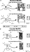
Figure 2. Number and distribution of Fos-IR single-labelled, GAD-IR single-labelled and Fos + GAD-IR double-labelled neurones located in counting grids for the rostral (A) and caudal (B) MnPN from one HSS rat and one LSS rat
In the HSS rat, 81% of rostral MnPN neurones and 77% of caudal MnPN neurones that stained for Fos-IR were also positive for GAD-IR. •, Fos + GAD double labelled; ▵, GAD single labelled; □, Fos single labelled
Figure 3
Figure 3. Mean numbers of total Fos-IR neurones, Fos-IR single-labelled neurones, Fos + GAD-IR double-labelled neurones and the mean percentage of Fos-IR neurones that were double labelled for GAD-IR in the rostral MnPN, caudal MnPN and vlPOA for HSS and LSS rats
**P < 0.001, independent t test; *P < 0.01, independent t test.
Figure 4
Figure 4. Regression lines and scatter plots of the number of Fos + GAD-IR double-labelled neurones versus total sleep time in the 11 rats studied in the spontaneous sleep protocol
In each of the three POA subregions examined, there was a significant positive correlation between sleep time and Fos + GAD-IR cell counts.
Figure 5
Figure 5. Temporal distribution of sleep–waking state progression (lower panels) and continuous EEG delta power (upper panels) during 2 h of recovery sleep in a sleep deprived rat and a sleep deprived control rat
Abbreviations: W, waking; N, non-REM sleep; R, REM sleep.
Figure 6
Figure 6. Mean numbers of Fos + GAD-IR double-labelled neurones (A) and mean percentage of GAD-IR neurones that were double labelled for Fos-IR (B) in SD rats permitted 2 h of recovery sleep (n= 6), in SDC rats permitted 2 h of recovery sleep (n= 6) and in HSS rats (n= 6)
ANOVA indicated a significant treatment effect on Fos + GAD-IR cell counts in the rostral MnPN (F2,15= 45.07, P < 0.001), the caudal MnPN (F2,15) = 27.78, P < 0.001) and the vlPOA (F2,15= 15.57, P < 0.001). Similarly, ANOVA indicated a significant treatment effect on the percentage of GAD-IR neurones that were double labelled for Fos-IR in the rostral MnPN (F2,15= 49.65, P < 0.001) caudal MnPN (F2,15= 14.29, P < 0.01) and vlPOA (F2,15= 13.08, P < 0.01). **Significantly different from SD and HSS values, P < 0.01, Newman–Keuls. *Significantly different from HSS values, P < 0.05, Newman–Keuls.
Figure 7
Figure 7. Mean numbers of Fos + GAD-IR double-labelled neurones in SD and SDC rats that were not permitted recovery sleep
The cell counts were significantly higher in SD versus SDC rats in the rostral MnPN (F1,10= 17.57, P < 0.01), the caudal MnPN (F1,10= 16.91, P < 0.01) and the vlPOA (F1,10= 15.57, P < 0.01).

References

    1. Alam MN, Gong H, Alam T, Jaganath R, McGinty D, Szymusiak R. Sleep-waking discharge patterns of neurons recorded in the rat perifornical lateral hypothalamic area. J Physiol. 2002;538:619–631. - PMC - PubMed
    1. Alam MN, McGinty D, Szymusiak R. Neuronal discharge of preoptic/anterior hypothalamic thermosensitive neurons: relation to NREM sleep. Am J Physiol. 1995a;269:R1240–R1249. - PubMed
    1. Alam MN, McGinty D, Szymusiak R. Thermosensitive neurons of the diagonal band in rats: relation to wakefulness and non-rapid eye movement sleep. Brain Res. 1997;752:81–89. - PubMed
    1. Alam N, Szymusiak R, McGinty D. Local preoptic/anterior hypothalamic warming alters spontaneous and evoked neuronal activity in the magno-cellular basal forebrain. Brain Res. 1995b;696:221–230. - PubMed
    1. Aston-Jones G, Bloom FE. Activity of norepinepherine-containing locus coeruleus neurons in behaving rats anticipates fluctuations in the sleep-waking cycle. J Neurosci. 1981;1:876–886. - PMC - PubMed

Publication types

MeSH terms

Substances

LinkOut - more resources