. 2004 Feb;113(4):539-50.
doi: 10.1172/JCI18829.

Inactivation of Icmt inhibits transformation by oncogenic K-Ras and B-Raf

Affiliations

Inactivation of Icmt inhibits transformation by oncogenic K-Ras and B-Raf

Martin O Bergo et al. J Clin Invest. 2004 Feb.

Abstract

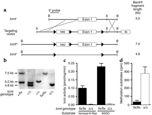
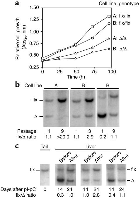
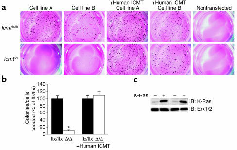
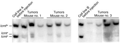
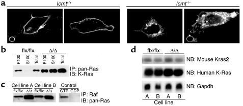
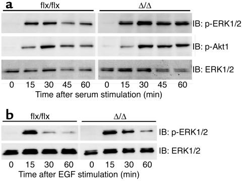
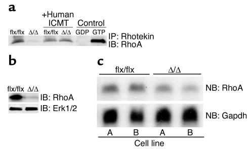
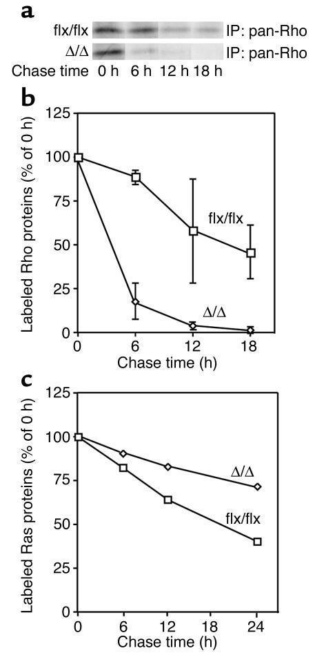
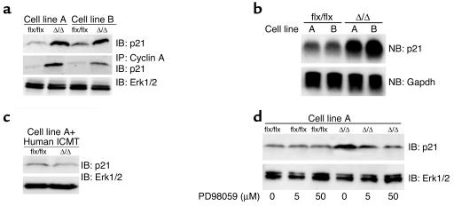
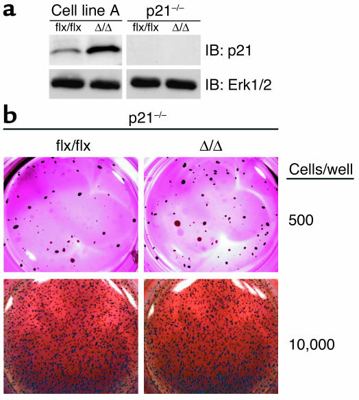
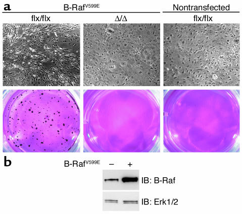
Isoprenylcysteine carboxyl methyltransferase (Icmt) methylates the carboxyl-terminal isoprenylcysteine of CAAX proteins (e.g., Ras and Rho proteins). In the case of the Ras proteins, carboxyl methylation is important for targeting of the proteins to the plasma membrane. We hypothesized that a knockout of Icmt would reduce the ability of cells to be transformed by K-Ras. Fibroblasts harboring a floxed Icmt allele and expressing activated K-Ras (K-Ras-Icmt(flx/flx)) were treated with Cre-adenovirus, producing K-Ras-Icmt(Delta/Delta) fibroblasts. Inactivation of Icmt inhibited cell growth and K-Ras-induced oncogenic transformation, both in soft agar assays and in a nude mice model. The inactivation of Icmt did not affect growth factor-stimulated phosphorylation of Erk1/2 or Akt1. However, levels of RhoA were greatly reduced as a consequence of accelerated protein turnover. In addition, there was a large Ras/Erk1/2-dependent increase in p21(Cip1), which was probably a consequence of the reduced levels of RhoA. Deletion of p21(Cip1) restored the ability of K-Ras-Icmt(Delta/Delta) fibroblasts to grow in soft agar. The effect of inactivating Icmt was not limited to the inhibition of K-Ras-induced transformation: inactivation of Icmt blocked transformation by an oncogenic form of B-Raf (V599E). These studies identify Icmt as a potential target for reducing the growth of K-Ras- and B-Raf-induced malignancies.


Figures

Figure 1
Figure 1
Generation and validation of a conditional Icmt allele (Icmtflx). (a) A sequence-replacement gene-targeting vector designed to flank exon 1 and upstream sequences with loxP sites. tk, thymidine kinase. (b) Southern blot identification of the Icmt+, Icmtflx, and IcmtΔ alleles with BamHI-cleaved genomic DNA and the 5′-flanking probe. (c) Icmt activity in extracts of Icmtflx/flx and IcmtΔ/Δ fibroblasts, as judged by a base-hydrolysis vapor-diffusion assay. Assays used S-adenosyl-L-[methyl-14C]methionine as the methyl donor and either farnesyl-K-Ras or N-acetyl-S-geranylgeranyl-L-cysteine (AGGC) as substrates. Bar graphs show the mean of two independent cell lines in two independent experiments. (d) Accumulation of Icmt substrates in IcmtΔ/Δ cells. Recombinant yeast Ste14p was added to extracts of Icmtflx/flx and IcmtΔ/Δ cells along with S-adenosyl-L-[methyl-14C]methionine; methylation of protein substrates was measured with the base-hydrolysis vapor-diffusion assay.
Figure 2
Figure 2
Analyzing the effect of Icmt inactivation on cell growth. (a) Anchorage-dependent growth of Icmtflx/flx and IcmtΔ/Δ cells. Equal numbers of immortalized Icmtflx/flx cells and the derivative IcmtΔ/Δ cells (lines A and B) were plated onto 96-well plates (n = 12 wells per cell line), and cell growth was assessed with the Cell Titer 96 AQueous One Solution Cell Proliferation Assay (Promega). (b) Southern blots illustrating the increase in the ratio of Icmtflx to IcmtΔ bands during the growth of a mixed population of Icmtflx/flx and IcmtΔ/Δ cells. Mixed populations of Icmtflx/flx and IcmtΔ/Δ cells were passaged at a 1:10 ratio every 3 days. DNA was harvested and analyzed with Southern blots at the indicated passages. (c) Southern blot of liver DNA from three Icmtflx/flxMx1-Cre mice before and after hepatocyte proliferation, which was induced by a partial hepatectomy. The ratio of Icmtflx to IcmtΔ band intensity increased after liver regrowth, indicating that Icmtflx/flx hepatocytes contributed more to liver regrowth than the IcmtΔ/Δ hepatocytes. Quantification of data from five mice revealed that the Icmtflx/IcmtΔ ratio increased 201% ± 26% after liver regeneration (P < 0.01).
Figure 3
Figure 3
Reduced capacity of K-Ras–transfected Icmt-deficient fibroblasts to form colonies in soft agar. (a) K-Ras-Icmtflx/flx and derivative K-Ras-IcmtΔ/Δ fibroblasts (2,000 cells of each) were mixed with 0.35% agarose and poured onto plates containing a 0.70% agarose base. Colonies were stained and photographed 21 days later. Nontransfected cells (i.e., no activated K-Ras) did not form colonies in soft agar. (b) Bar graph illustrating the number of colonies formed in soft agar in four independent experiments; data in each experiment were normalized to the number of colonies that formed with the parental K-Ras-Icmtflx/flx fibroblasts. Inactivation of Icmt significantly reduced the number of colonies that formed in soft agar (*P < 0.0001). For experiments involving K-Ras-Icmtflx/flx:ICMT cells (expressing a human ICMT cDNA) and the derivative K-Ras-IcmtΔ/Δ:ICMT cells, data show the results from three independent experiments. In the cells expressing human ICMT, inactivation of mouse Icmt did not affect colony formation (P = 0.63). (c) Western blot showing higher K-Ras expression levels in K–Ras-transfected cells (+) compared with nontransfected cells (–). The blot was stripped and incubated with an anti-Erk1/2 antibody as a loading control. IB, immunoblot.
Figure 4
Figure 4
Impaired ability of K-Ras–transfected Icmt-deficient fibroblasts to contribute to tumor growth in nude mice. A mixed population of K-Ras-Icmtflx/flx cells and derivative K-Ras-IcmtΔ/Δ cells (total of 500,000 cells, prepared as described in the Methods section) was injected subcutaneously into nude mice. Tumors were harvested 21 days later. Southern blots were performed on genomic DNA prepared from the injected cells and the tumors. The ratio of Icmtflx to IcmtΔ band intensity was much greater in the tumor DNA than in the injected cells. A 5-kb Icmt+ allele in some lanes was due to host DNA in the tumors.
Figure 5
Figure 5
Mislocalization and increased steady-state levels of K-Ras in Icmt-deficient fibroblasts. (a) Confocal micrographs of spontaneously immortalized Icmt+/+ and Icmt–/– fibroblasts that had been transfected with a GFP–K-Ras fusion construct. (b) Distribution of Ras proteins in the membrane (P100) and cytosolic (S100) fractions of K-Ras-Icmtflx/flx fibroblasts and the derivative K-Ras-IcmtΔ/Δ fibroblasts. The Ras proteins were immunoprecipitated from 1,000 μg of the indicated cell fractions with a pan-Ras–specific antibody, and a Western blot was performed with a K-Ras–specific antibody. (c) GTP-bound Ras proteins in K-Ras-Icmtflx/flx fibroblasts and derivative K-Ras-IcmtΔ/Δ fibroblasts. GTP-bound Ras proteins were precipitated from 1 × 10⁶ K-Ras-Icmtflx/flx and K-Ras-IcmtΔ/Δ cells with the Ras-binding domain of Raf (Ras Activation Kit; Upstate Biotechnology Inc.), and a Western blot was performed with a pan-Ras antibody. (d) Northern blot of total RNA from K-Ras-Icmtflx/flx and K-Ras-IcmtΔ/Δ fibroblasts, hybridized with a mouse Kras2 cDNA probe. That probe detects a longer mouse Kras2 transcript (upper panel) and a shorter human activated K-Ras transcript (middle panel). The blot was stripped and hybridized with a Gapdh cDNA probe (lower panel). IP, immunoprecipitation; NB, Northern blot.
Figure 6
Figure 6
Growth factor-stimulated Erk1/2 and Akt1 phosphorylation in Icmt-deficient fibroblasts. (a) Nontransfected Icmtflx/flx and the derivative IcmtΔ/Δ fibroblasts were seeded at equal density and serum-starved overnight as described in the Methods section. Serum-containing medium was then added to the cells. Cells were harvested at the indicated time points and analyzed by immunoblotting with antibodies against phosphorylated Erk1/2 (p-Erk1/2), phosphorylated Akt1 (p-Akt1), and total Erk1/2. (b) Icmtflx/flx and the derivative IcmtΔ/Δ fibroblasts were seeded at equal density and serum-starved overnight. Medium (0.5% serum) supplemented with EGF (50 ng/ml) was added to the cells. Cells were harvested at the indicated time points and analyzed by immunoblotting with antibodies against p-Erk1/2 and total Erk1/2.
Figure 7
Figure 7
Low steady-state levels of RhoA in Icmt-deficient fibroblasts. (a) GTP-bound Rho proteins were immunoprecipitated from 1 × 10⁶ K-Ras-Icmtflx/flx fibroblasts and derivative K-Ras-IcmtΔ/Δ fibroblasts with equal amounts of Rhotekin-GST (Rho Activation Kit; Upstate Biotechnology Inc.). The GTP-bound Rho proteins were resolved by SDS-PAGE and detected with a RhoA-specific antibody (26C4 monoclonal; Santa Cruz Biotechnology Inc.). In the same experiment, bands for two control proteins were of identical intensity in the K-Ras-Icmtflx/flx and K-Ras-IcmtΔ/Δ fibroblasts (not shown). Similar results were obtained when using a pan-Rho antibody. (b) Cell extracts from K-Ras-Icmtflx/flx fibroblasts and the derivative K-Ras-IcmtΔ/Δ fibroblasts were analyzed by immunoblotting with a RhoA-specific antibody. The blot was stripped and incubated with an anti-Erk1/2 antibody as a loading control. (c) Northern blot (NB) of total cellular RNA from K-Ras-Icmtflx/flx fibroblasts and the derivative K-Ras-IcmtΔ/Δ fibroblasts was hybridized with a mouse RhoA cDNA probe. Reprobing of the membrane with a Gapdh cDNA probe revealed similar levels of Gapdh expression in each sample (see Figure 5d).
Figure 8
Figure 8
Accelerated turnover of Rho proteins in Icmt-deficient fibroblasts. (a) K-Ras-Icmtflx/flx and K-Ras-IcmtΔ/Δ fibroblasts were labeled with [35S]methionine/cysteine for 2 hours and then incubated in regular medium supplemented with 4 mM “cold” methionine and cysteine for the indicated times. Rho proteins were immunoprecipitated from total cell extracts with a pan-Rho antibody, resolved by SDS-PAGE, and then visualized by autoradiography as described in the Methods section. (b) Pulse-chase analysis of Rho protein disappearance in K-Ras-Icmtflx/flx and the derivative K-Ras-IcmtΔ/Δ fibroblasts. Graphs show mean densitometry results from three independent experiments. (c) Pulse-chase analysis of Ras proteins in K-Ras-Icmtflx/flx and the derivative K-Ras-IcmtΔ/Δ fibroblasts. Ras proteins were immunoprecipitated from total cell extracts with a pan-Ras antibody, resolved by SDS-PAGE, and then visualized by autoradiography. Graphs show mean densitometry data from two independent experiments.
Figure 9
Figure 9
Increased p21Cip1 protein levels in Icmt-deficient fibroblasts. (a) Extracts from K-Ras-Icmtflx/flx and the derivative K-Ras-IcmtΔ/Δ fibroblasts were analyzed by immunoblotting with an antibody recognizing p21Cip1 (F-5 monoclonal; Santa Cruz Biotechnology Inc.) (upper panel). Cyclin A was immunoprecipitated from the cell extracts with a polyclonal antibody (H-432; Santa Cruz Biotechnology Inc.), and cyclin A–associated p21Cip1 was detected by immunoblotting (middle panel). The blot from the upper panel was stripped and incubated with an anti-Erk1/2 antibody as a loading control (lower panel). Similar results were obtained in three independent experiments. (b) Northern blot of total cellular RNA showing p21Cip1 (Cdkn1a) mRNA levels in K-Ras-Icmtflx/flx fibroblasts and the derivative K-Ras-IcmtΔ/Δ fibroblasts (upper panel). The blot was stripped and probed with a Gapdh cDNA probe as a loading control (lower panel). Similar results were obtained in three independent experiments. (c) Immunoblot showing p21Cip1 protein levels in K-Ras-Icmtflx/flx:ICMT fibroblasts and the derivative K-Ras-IcmtΔ/Δ:ICMT fibroblasts. The blot was stripped and incubated with an anti-Erk1/2 antibody as a loading control (lower panel). (d) K-Ras-Icmtflx/flx and K-Ras-IcmtΔ/Δ fibroblasts were treated overnight with the MEK inhibitor PD98059, and extracts were analyzed by immunoblotting with a p21Cip1-specific antibody. The blot was stripped and incubated with an anti-Erk1/2 antibody as a loading control (lower panel).
Figure 10
Figure 10
Knocking out p21Cip1 restores the capacity of K-Ras-IcmtΔ/Δ fibroblasts to grow in soft agar. (a) Western blot showing the absence of p21Cip1 protein in K-Ras-Icmtflx/flxCdkn1a–/– fibroblasts and the derivative K-Ras-IcmtΔ/ΔCdkn1a–/– fibroblasts. The blot was stripped and incubated with an anti-Erk1/2 antibody as a loading control (lower panel). (b) K-Ras-Icmtflx/flxCdkn1a–/– and K-Ras-IcmtΔ/ΔCdkn1a–/– fibroblasts were seeded in soft agar. After 30 days, the colonies were stained, photographed, and analyzed as described in the Methods section. Similar results were obtained in three independent experiments; there was no difference in colony numbers with K-Ras-Icmtflx/flxCdkn1a–/– and the derivative K-Ras-IcmtΔ/ΔCdkn1a–/– fibroblasts (P = 0.47).
Figure 11
Figure 11
Inactivation of Icmt reverses B-Raf–induced transformation. (a) B-Raf-Icmtflx/flx and B-Raf-IcmtΔ/Δ fibroblasts were grown to confluence and then allowed to grow for an additional 6 days. The cells were then fixed in 4% paraformaldehyde and photographed (upper panel). An aliquot of the same cells was seeded in soft agar (lower panel). After 20 days, the colonies were stained, photographed, and analyzed as described in the Methods section. Similar results were obtained in three independent experiments. Transfection of fibroblasts with a retrovirus encoding wild-type B-Raf did not yield a transformed phenotype (not shown). (b) Western blot with a B-Raf–specific mAb (F-7; Santa Cruz Biotechnology Inc.) showing increased B-Raf expression in Icmtflx/flx fibroblasts transfected with the retrovirus encoding human B-RafV599E. The blot was stripped and incubated with an anti-Erk1/2 antibody as a loading control (lower panel).

Comment in

References

    1. Casey PJ, Seabra MC. Protein prenyltransferases. J. Biol. Chem. 1996;271:5289–5292. - PubMed
    1. Boyartchuk VL, Ashby MN, Rine J. Modulation of Ras and a-factor function by carboxyl-terminal proteolysis. Science. 1997;275:1796–1800. - PubMed
    1. Dai Q, et al. Mammalian prenylcysteine carboxyl methyltransferase is in the endoplasmic reticulum. J. Biol. Chem. 1998;273:15030–15034. - PubMed
    1. Goodman LE, et al. Mutants of Saccharomyces cerevisiae defective in the farnesylation of Ras proteins. Proc. Natl. Acad. Sci. U. S. A. 1990;87:9665–9669. - PMC - PubMed
    1. Reiss Y, Goldstein JL, Seabra MC, Casey PJ, Brown MS. Inhibition of purified p21ras farnesyl:protein transferase by Cys-AAX tetrapeptides. Cell. 1990;62:81–88. - PubMed

Publication types

MeSH terms