. 2004 Feb;113(4):551-60.
doi: 10.1172/JCI19133.

Inhibition of airway remodeling in IL-5-deficient mice

Affiliations

Inhibition of airway remodeling in IL-5-deficient mice

Jae Youn Cho et al. J Clin Invest. 2004 Feb.

Abstract

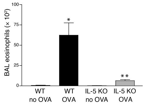
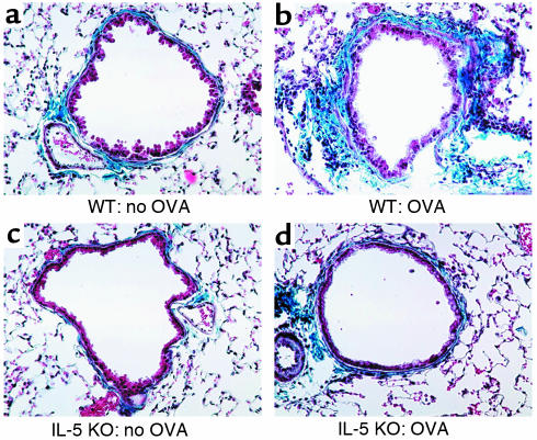
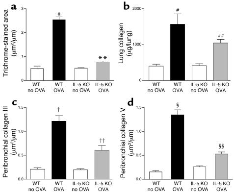
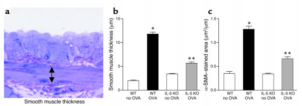
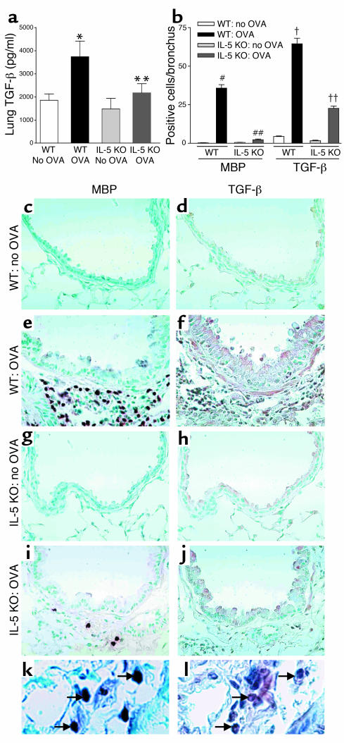
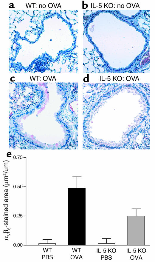
To determine the role of IL-5 in airway remodeling, IL-5-deficient and WT mice were sensitized to OVA and challenged by repetitive administration of OVA for 3 months. IL-5-deficient mice had significantly less peribronchial fibrosis (total lung collagen content, peribronchial collagens III and V) and significantly less peribronchial smooth muscle (thickness of peribronchial smooth muscle layer, alpha-smooth muscle actin immunostaining) compared with WT mice challenged with OVA. WT mice had a significant increase in the number of peribronchial cells staining positive for major basic protein and TGF-beta. In contrast, IL-5-deficient mice had a significant reduction in the number of peribronchial cells staining positive for major basic protein, which was paralleled by a similar reduction in the number of cells staining positive for TGF-beta, suggesting that eosinophils are a significant source of TGF-beta in the remodeled airway. OVA challenge induced significantly higher levels of airway epithelial alphaVbeta6 integrin expression, as well as significantly higher levels of bioactive lung TGF-beta in WT compared with IL-5-deficient mice. Increased airway epithelial expression of alphaVbeta6 integrin may contribute to the increased activation of latent TGF-beta. These results suggest an important role for IL-5, eosinophils, alphaVbeta6, and TGF-beta in airway remodeling.


Figures

Figure 1
Figure 1
BALF eosinophil levels in WT and IL-5–deficient mice repetitively challenged with OVA. WT mice repetitively challenged with OVA for 3 months developed significantly increased numbers of BALF eosinophils compared with non–OVA-challenged WT mice (*WT OVA versus WT no OVA; P < 0.001). In contrast IL-5–deficient mice repetitively challenged with OVA for 3 months had significantly reduced levels of BALF eosinophils compared with WT mice repetitively challenged with OVA for 3 months (**IL-5 KO OVA versus WT OVA; P < 0.01).
Figure 2
Figure 2
Peribronchial trichrome stain in WT and IL-5–deficient mice repetitively challenged with OVA. In the absence of OVA challenge, WT (a) and IL-5–deficient mice (c) exhibited minimal peribronchial trichrome staining (blue color). In contrast, repetitive OVA challenge in WT mice for 3 months induced circumferential peribronchial trichrome staining (b), which was significantly reduced in IL-5–deficient mice repetitively challenged with OVA (d).
Figure 3
Figure 3
Quantitation of peribronchial fibrosis in WT and IL-5–deficient mice repetitively challenged with OVA. (a) Area of peribronchial trichrome stain. WT mice repetitively challenged with OVA for 3 months developed an increased area of peribronchial trichrome staining compared with non–OVA-challenged WT mice (*WT OVA versus WT no OVA; P < 0.001). In contrast, IL-5–deficient mice repetitively challenged with OVA for 3 months had significantly reduced areas of peribronchial trichrome staining compared with WT mice repetitively challenged with OVA for 3 months (**IL-5 KO OVA versus WT OVA; P < 0.001). (b) Total lung collagen content. Repetitive OVA challenge induced a significant increase in total lung collagen in WT mice (#WT OVA versus WT no OVA; P < 0.001). IL-5–deficient mice repetitively challenged with OVA had less total lung collagen compared with WT mice repetitively challenged with OVA (##IL-5 KO OVA versus WT OVA; P < 0.05). (c) Collagen III and (d) collagen V lung immunostaining. WT mice repetitively challenged with OVA for 3 months developed increased peribronchial collagen III immunostaining (WT OVA versus WT no OVA; P < 0.001) (c), as well as increased peribronchial collagen V immunostaining compared with non-OVA–challenged WT mice (§WT OVA versus WT no OVA; P < 0.001) (d). In contrast, IL-5–deficient mice repetitively challenged with OVA had significantly reduced levels of peribronchial collagen III immunostaining (††IL-5 KO OVA versus WT OVA; P < 0.001) (c), as well as significantly reduced levels of peribronchial collagen V immunostaining, compared with WT mice challenged repetitively with OVA for 3 months (§§IL-5 KO OVA versus WT OVA; P < 0.001) (d).
Figure 4
Figure 4
Peribronchial smooth muscle layer in WT and IL-5–deficient mice repetitively challenged with OVA. (a) Peribronchial smooth muscle layer. Lungs that had been fixed in 3% glutaraldehyde and 1% osmium tetroxide were stained with basic fuchsin-toluidine blue, which allowed the best visualization of the peribronchial smooth muscle layer. The thickness of the smooth muscle layer (the transverse diameter) was measured from the innermost aspect to the outermost aspect of the smooth muscle layer (double arrow). (b) Peribronchial smooth muscle layer thickness. WT mice repetitively challenged with OVA for 3 months developed significantly increased thickness of the peribronchial smooth muscle layer compared with non–OVA-challenged WT mice (*WT OVA versus WT no OVA; P < 0.001). In contrast, the thickness of the peribronchial smooth muscle layer in IL-5–deficient mice challenged repetitively with OVA was significantly reduced compared with WT mice challenged repetitively with OVA for 3 months (**IL-5 KO OVA versus WT OVA; P < 0.001). (c) Peribronchial α-smooth muscle actin (α-SMA). WT mice repetitively challenged with OVA for 3 months developed an increase in the area of peribronchial α-smooth muscle actin immunostaining compared with non–OVA-challenged WT mice (*WT OVA versus WT no OVA; P < 0.001). The area of peribronchial α-smooth muscle actin immunostaining in IL-5–deficient mice challenged repetitively with OVA was significantly reduced compared with WT mice challenged repetitively with OVA for 3 months (**IL-5 KO OVA versus WT OVA; P < 0.001).
Figure 5
Figure 5
Lung TGF-β1 in WT and IL-5–deficient mice repetitively challenged with OVA. (a) WT mice challenged with OVA developed increased levels of lung TGF-β1 compared with non–OVA-challenged WT mice (*WT OVA versus WT no OVA; P < 0.01). Significantly reduced levels of TGF-β1 were detected in IL-5–deficient mice versus WT mice challenged with OVA (**IL-5 KO OVA versus WT OVA; P < 0.05). (b) WT mice challenged with OVA had a significant increase in the number of peribronchial MBP-positive cells (#WT OVA versus WT no OVA; P < 0.001) and TGF-β–positive cells (WT OVA versus WT no OVA; P < 0.001). There was a significant reduction in the number of MBP-positive cells (##IL-5 KO OVA versus WT OVA; P < 0.001) and TGF-β–positive cells (††IL-5 KO OVA versus WT OVA; P < 0.001) in IL-5–deficient mice challenged with OVA. (cj) Non–OVA-challenged WT mice had few cells that immunostained positive for MBP (c) or TGF-β (d). OVA-challenged WT mice had a significant increase in the number of cells that immunostain positive for MBP (e) or TGF-β (f). OVA challenged IL-5–deficient mice had few cells that immunostained positive for MBP (i) or TGF-β (j). Epithelial cells in OVA-challenged IL-5–deficient mice did immunostain positive for TGF-β (j). Non–OVA-challenged IL-5–deficient mice had few cells that immunostained positive for MBP (g) or TGF-β (h). The arrows in k point to three of four MBP-positive cells. The arrows in l point to three cells that are TGF-β1 positive (corresponding to the same three MBP-positive cells in k). Several additional TGF-β1–positive cells that do not correspond to MBP-positive cells are also noted in l.
Figure 6
Figure 6
Expression of airway epithelial integrin αVβ6 in WT and IL-5 KO mice. Neither non–OVA-challenged WT mice (a) nor non–OVA-challenged IL-5–deficient mice (b) had significant numbers of airway epithelial cells that immunostained positive for αVβ6. In contrast, WT mice challenged repetitively with OVA for 3 months had a significant increase in the number of airway epithelial cells that immunostained positive for αVβ6 (c). There was a significant reduction in IL-5–deficient mice in the number of airway epithelial cells that immunostain positive for αVβ6 following repetitive OVA challenge (d). Quantitative analysis of αVβ6 integrin expression in airway epithelial cells demonstrated that repetitive OVA challenge in WT mice induced a significant increase in αVβ6 expression in WT mice compared with diluent-challenged WT mice (WT OVA versus WT no OVA; P < 0.001). The area of airway epithelium staining positively for αVβ6 in repetitively OVA-challenged IL-5–deficient mice was significantly less than that of repetitively OVA-challenged WT mice (IL-5 KO OVA versus WT OVA) (P < 0.001) (e).

Comment in

  • The eosinophil enigma.
    Williams TJ. Williams TJ. J Clin Invest. 2004 Feb;113(4):507-9. doi: 10.1172/JCI21073. J Clin Invest. 2004. PMID: 14966558 Free PMC article.

References

    1. Rothenberg ME. Eosinophilia. N. Engl. J. Med. 1998;338:1592–1600. - PubMed
    1. Broide D. Fast flowing eosinophils: signals for stopping and stepping out of blood vessels. Am. J. Respir. Cell Mol. Biol. 2002;26:637–640. - PubMed
    1. Lopez AF, et al. Recombinant human interleukin 5 is a selective activator of human eosinophil function. J. Exp. Med. 1988;167:219–224. - PMC - PubMed
    1. Foster PS, Hogan SP, Ramsay AJ, Matthaei KI, Young IG. Interleukin 5 deficiency abolishes eosinophilia, airways hyperreactivity, and lung damage in a mouse asthma model. J. Exp. Med. 1996;183:195–201. - PMC - PubMed
    1. Lee JJ, et al. Interleukin-5 expression in the lung epithelium of transgenic mice leads to pulmonary changes pathognomonic of asthma. J. Exp. Med. 1997;185:2143–2156. - PMC - PubMed

Publication types

MeSH terms