. 2004 Feb;113(4):561-8.
doi: 10.1172/JCI19081.

Targeted ablation of Fgf23 demonstrates an essential physiological role of FGF23 in phosphate and vitamin D metabolism

Affiliations

Targeted ablation of Fgf23 demonstrates an essential physiological role of FGF23 in phosphate and vitamin D metabolism

Takashi Shimada et al. J Clin Invest. 2004 Feb.

Abstract

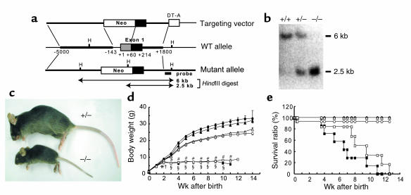
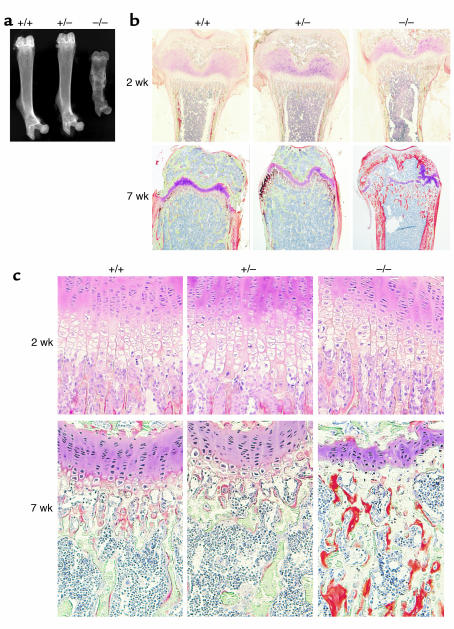
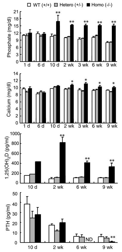
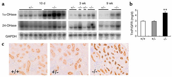
Inorganic phosphate is essential for ECM mineralization and also as a constituent of important molecules in cellular metabolism. Investigations of several hypophosphatemic diseases indicated that a hormone-like molecule probably regulates serum phosphate concentration. FGF23 has recently been recognized as playing important pathophysiological roles in several hypophosphatemic diseases. We present here the evidence that FGF23 is a physiological regulator of serum phosphate and 1,25-dihydroxyvitamin D (1,25[OH]2D) by generating FGF23-null mice. Disruption of the Fgf23 gene did not result in embryonic lethality, although homozygous mice showed severe growth retardation with abnormal bone phenotype and markedly short life span. The Fgf23(-/-) mice displayed significantly high serum phosphate with increased renal phosphate reabsorption. They also showed an elevation in serum 1,25(OH)2D that was due to the enhanced expression of renal 25-hydroxyvitamin D-1alpha-hydroxylase (1alpha-OHase) from 10 days of age. These phenotypes could not be explained by currently known regulators of mineral homeostasis, indicating that FGF23 is essential for normal phosphate and vitamin D metabolism.


Figures

Figure 1
Figure 1
Establishment of FGF23-null mice. (a) Targeted ablation of Fgf23 gene. Construct of a targeting vector (top), wild-type allele (middle), and targeted mutant allele (bottom) are shown. DT-A, diphtheria toxin-A fragment; H, HindIII. (b) Genomic Southern blot analysis. Genomic DNAs (5 μg) isolated from transfected ES clones were digested with HindIII and hybridized to the indicated probe. (c) Gross phenotypes of heterozygous and homozygous founders at 6 weeks of age. (d) Growth curves and (e) survival ratios for male (filled) or female (open) WT (circles), heterozygotes (triangles), and homozygotes (squares). Data are represented as mean ± SEM. Statistical analysis in each sex was carried out by Dunnett’s method. Statistically significant results are marked by asterisk (male) or cross (female) (*P < 0.05, §P < 0.01, P < 0.05, #P < 0.01). Numbers of animals used in this study: male WT (+/+), n = 9; female WT, n = 15; male heterozygotes (+/–), n = 20; female heterozygotes, n = 19; male homozygotes (–/–), n = 7; female homozygotes, n = 6.
Figure 2
Figure 2
Histological analysis of bone. (a) Soft X-rays of femurs prepared from 7-week-old mice. (b and c) Villanueva-Goldner stain of undecalcified femurs prepared from 2- and 7-week-old mice. (b) Distal metaphysis of femurs isolated at 2 weeks (upper) and 7 weeks (lower) of age. Original magnification, ×4. (c) Growth plates of femurs at 2 weeks (upper) and 7 weeks (lower) of age. Mineralized bone tissues are colored in green, and unmineralized osteoid tissues are shown in orange. Original magnification, ×20.
Figure 3
Figure 3
Comparison of serum parameters. Blood samples were collected from carotid artery (day 1 and 6) or heart (day 10 and thereafter) under conditions of anesthesia. Because sufficient amounts of blood could not be obtained from mice aged younger than 10 days, sera were pooled in each genotype for measurement of 1,25(OH)2D. Other results represent mean ± SEM. The numbers of mice killed at each age were variable (average; WT n = 5, heterozygotes n = 9, and homozygotes n = 6). Statistical analysis was carried out by Dunnett’s method for comparison of multiple means (*P < 0.05; **P < 0.01). ND, not detected.
Figure 4
Figure 4
Key molecules for serum phosphate and 1,25(OH)2D levels. (a) Northern blot analysis of renal 1α-OHase and 24-OHase mRNAs. The blots were reprobed with a GAPDH fragment to confirm integrity of the electrophoresed RNAs. (b) TmP/GFR. Mice (6 weeks old) were reared in metabolic cages to collect urine samples for 24 hours. The results represent mean ± SEM. WT, n = 6; heterozygotes, n = 6; homozygotes, n = 6. **P < 0.01, evaluated by Dunnett’s method. (c) Immunohistochemistry of renal NaPi-2a protein at 6 weeks of age.

References

    1. Brown EM, et al. Cloning and characterization of an extracellular Ca(2+)-sensing receptor from bovine parathyroid. Nature. 1993;366:575–580. - PubMed
    1. Fitzpatrick, L.A., and Bilezikian, J.P. 1996. Actions of parathyroid hormone. In Principles of bone biology. J.P. Bilezikian, L.G. Raisz, and G.A. Rodan, editors. Academic Press Inc. San Diego, California, USA. 339–346.
    1. Drezner MK. PHEX gene and hypophosphatemia. Kidney Int. 2000;57:9–18. - PubMed
    1. Econs MJ, Drezner MK. Tumor-induced osteomalacia—unveiling a new hormone. N. Engl. J. Med. 1994;330:1679–1681. - PubMed
    1. The ADHR Consortium. Autosomal dominant hypophosphataemic rickets is associated with mutations in FGF23. Nat. Genet. 2000;26:345–348. - PubMed

Publication types