. 2004 Feb;113(4):598-608.
doi: 10.1172/JCI18776.

The calcium-sensing receptor regulates mammary gland parathyroid hormone-related protein production and calcium transport

Affiliations

The calcium-sensing receptor regulates mammary gland parathyroid hormone-related protein production and calcium transport

Joshua VanHouten et al. J Clin Invest. 2004 Feb.

Abstract

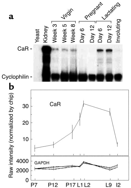
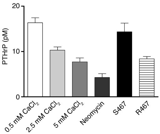
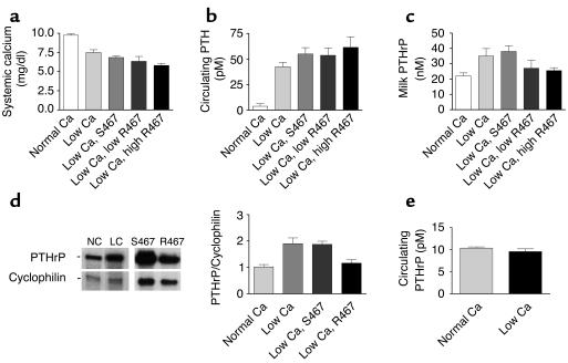
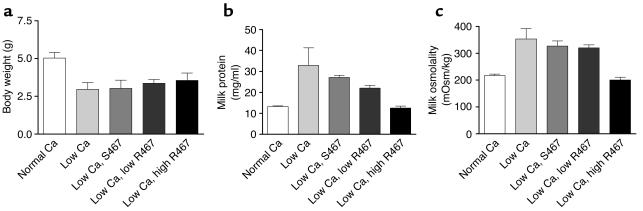
The transfer of calcium from mother to milk during lactation is poorly understood. In this report, we demonstrate that parathyroid hormone-related protein (PTHrP) production and calcium transport in mammary epithelial cells are regulated by extracellular calcium acting through the calcium-sensing receptor (CaR). The CaR becomes expressed on mammary epithelial cells at the transition from pregnancy to lactation. Increasing concentrations of calcium, neomycin, and a calcimimetic compound suppress PTHrP secretion by mammary epithelial cells in vitro, whereas in vivo, systemic hypocalcemia increases PTHrP production, an effect that can be prevented by treatment with a calcimimetic. Hypocalcemia also reduces overall milk production and calcium content, while increasing milk osmolality and protein concentrations. The changes in milk calcium content, milk osmolality, and milk protein concentration were mitigated by calcimimetic infusions. Finally, in a three-dimensional culture system that recapitulates the lactating alveolus, activation of the basolateral CaR increases transcellular calcium transport independent of its effect on PTHrP. We conclude that the lactating mammary gland can sense calcium and adjusts its secretion of calcium, PTHrP, and perhaps water in response to changes in extracellular calcium concentration. We believe this defines a homeostatic system that helps to match milk production to the availability of calcium.


Figures

Figure 1
Figure 1
CaR expression over the course of mammary gland development. (a) RNase protection analysis of CaR mRNA levels during mouse mammary gland development. Fifty micrograms of total RNA prepared from mouse mammary glands harvested at the timepoints noted in the figure was assayed. Five micrograms of mouse kidney RNA served as a positive control. Fifty micrograms of yeast RNA served as a negative control. (b) Profile of CaR transcript representation in an Affymetrix gene expression database. Each point represents the mean of four separate RNA samples prepared from mammary glands harvested at the indicated times. Error bars represent the SEM. P7, P12, and P17 represent days 7, 12, and 17 of pregnancy, respectively; L1, L2, and L9 represent days 1, 2, and 9 of lactation, respectively; I2 represents the second day of involution after weaning. The profile for GAPDH transcripts is provided for comparison.
Figure 2
Figure 2
Immunofluorescence staining for the CaR in mammary glands of lactating mice (a and b) and in mammospheres (c and d). Staining was done with 10 μg/ml anti-CaR antibody (a and c) or rabbit IgG (b and d), and images were produced using a Zeiss LSM 510 confocal microscope. Arrowheads in a indicate basolateral membrane staining. Scale bars: 20 μm.
Figure 3
Figure 3
PTHrP concentrations in conditioned media harvested from cultures of normal mammary epithelial cells exposed to the various concentrations of calcium noted on the graph. Cells were also exposed to 300 μM of neomycin and 2.5 μM of the calcimimetic NPS R467 or its less active isomer NPS S467. Bars represent the mean of three experiments; error bars represent the SEM. The differences between 0.5 mM and 2.5 mM or 5.0 mM CaCl2 were significant (P < 0.01 for 0.5 mM vs. 2.5 mM, P < 0.001 for 0.5 mM vs. 5.0 mM), but the difference between 2.5 and 5.0 mM was not. The differences between 0.5 mM CaCl2 and neomycin were significant (P < 0.001) as were the differences between NPS S467 and NPS R467 (P < 0.01).
Figure 4
Figure 4
Plasma calcium, PTH, and PTHrP levels, milk PTHrP concentrations, and PTHrP mRNA expression levels in lactating mice fed a normal calcium diet, a low calcium diet, or a low calcium diet and treated with either NPS S467 or NPS R467. (a) Plasma calcium concentrations in mice fed either a normal-calcium diet (0.6%) or a low-calcium diet (0.01%). Mice on a low-calcium diet either received nothing (low Ca) or were infused with NPS S467 at 40 μmol/kg/d or with NPS R467 at doses of 4 μmol/kg/d (low R467) or 40 μmol/kg/d (high R467). Calcium restriction led to a significant decline in plasma calcium (P < 0.001, normal Ca vs. low Ca). The higher dose of NPS R467 led to a further significant decline in plasma calcium (P < 0.01, low Ca vs. low Ca plus high R467). (b) Plasma PTH in mice on normal or low-calcium diets with or without calcimimetic treatment. (c) Milk PTHrP concentrations in the same groups described above. Milk PTHrP concentrations were significantly different in groups given low Ca (P < 0.05) and low Ca plus S467 (P < 0.05) compared with the group on a normal diet. However, milk PTHrP concentrations in the mice treated with NPS R467 were not significantly different from baseline. (d) PTHrP mRNA levels in mammary glands of mice on a normal-calcium diet (NC), a low-calcium diet (LC), a low-calcium diet with NPS S467 treatment (40 μmol/kg/d), or a low-calcium diet with NPS R467 treatment (40 μmol/kg/d) assessed by RNase protection analysis. Forty micrograms of total RNA was assayed. The bar graph represents cumulative data from four animals per treatment normalized to cyclophilin. (e) Circulating plasma PTHrP levels in mice fed a normal-calcium diet or a low-calcium diet.
Figure 5
Figure 5
Pup weight, milk protein concentration, and milk osmolality measured in lactating dams receiving a normal calcium diet or in calcium-restricted lactating mice treated with a calcimimetic or the control compound. (a) Average pup body weight on day 12 of lactation in litters suckling on dams fed a normal-calcium diet or a low-calcium diet with or without calcimimetic treatment. Mice received 40 μmol/kg/d of NPS S467 or one of two doses of NPS R467, 4 μmol/kg/d (low) or 40 μmol/kg/d (high). Pups were significantly smaller when mothers consumed a low-calcium diet. Calcimimetic treatment did not significantly increase pup weight. (b) Protein concentration of milk derived from the groups of mice described in a. Each bar represents the mean of three to nine samples; error bars represent the SEM. Milk protein was significantly higher in samples from mice on a low-calcium diet compared with mice on a normal-calcium diet (P < 0.001) or mice receiving NPS R467 (P < 0.01). However, the protein concentrations were not significantly different in milk from mice receiving NPS R467 compared with milk from those on a normal diet. (c) Milk osmolality in samples from the same groups described in a and b. Osmolality was significantly increased in milk from calcium-restricted mice (P < 0.001 vs. normal Ca) and mice receiving NPS S467 (P < 0.01 vs. normal Ca). The low dose of NPS R467 did not significantly decrease milk osmolality, but the high dose did (P < 0.001 for low Ca vs. low Ca plus high R467). mOsm, milliosmoles.
Figure 6
Figure 6
Milk calcium content corrected for milk protein concentration in samples from mice fed either a normal-calcium diet or a low-calcium diet and either treated with NPS S467 or NPS R467 at two different doses. Bars represent the means of three to nine samples; error bars represent the SEM. Calcium restriction led to a significant decrease in the calcium content of the milk (P < 0.001 for normal Ca vs. low Ca). Treatment with the higher dose of NPS R467 significantly increased the calcium content of the milk compared with a low-calcium diet (P < 0.05); however it remained significantly lower than the calcium content in mice fed a normal diet (P < 0.05).
Figure 7
Figure 7
CaR-stimulated transcellular calcium transport in mouse mammary epithelial cells cultured on Matrigel. (a) Fluorescent micrograph of a section through a mammosphere incubated with NHS-LC-biotin and stained with fluorescein-tagged avidin. As one can see, NHS-LC-biotin added to the media is excluded from the lumen, documenting that there is no paracellular leak through the tight junctions between epithelial cells. Lu, lumen; am, apical membrane; bm, basolateral membrane. (b) Same as in a, except that in addition to NHS-LC-biotin, 2.5 mM EGTA was added to the media of the mammosphere cultures. In this instance, the tight junctions became leaky and the NHS-LC-biotin labeled both basolateral and apical membranes. (c) 45Ca accumulation within the lumens of mammospheres made from WT mice (white bars) or from BLG-Cre/PTHrPlox/– mice (black bars) cultured in 1, 5, or 10 mM CaCl2 or in 1 mM CaCl2 with 2.5 μM NPS S467 or NPS R467 added. Mammary epithelial cells from the BLG-Cre/PTHrPlox/– mice did not secrete PTHrP (not shown). As can be seen, extracellular calcium stimulates the accumulation of 45Ca in the lumen of mammospheres in a dose-dependent manner, regardless of the presence or absence of PTHrP. Likewise, stimulation of CaR signaling with NPS R467 led to a significant increase in the luminal accumulation of tracer compared with treatment with NPS S467 in both types of cells (WT, P < 0.0001; BLG-Cre/PTHrPlox–, P < 0.05). Each bar represents the mean of three experiments; error bars represent the SEM.

References

    1. Brown EM, et al. Cloning and characterization of an extracellular Ca(2+)-sensing receptor from bovine parathyroid. Nature. 1993;366:575–580. - PubMed
    1. Brown EM, MacLeod RJ. Extracellular calcium sensing and extracellular calcium signaling. Physiol. Rev. 2001;81:239–297. - PubMed
    1. Chattopadhyay N, Mithal A, Brown EM. The calcium-sensing receptor: a window into the physiology and pathophysiology of mineral ion metabolism. Endocr. Rev. 1996;17:289–307. - PubMed
    1. Brown EM. Extracellular Ca2+ sensing, regulation of parathyroid cell function, and role of Ca2+ and other ions as extracellular (first) messengers. Physiol. Rev. 1991;71:371–411. - PubMed
    1. Conklin BR, Bourne HR. Homeostatic signals. Marriage of the flytrap and the serpent. Nature. 1994;367:22. - PubMed

Publication types

MeSH terms