Review
. 2004 Feb;37(2):79-85.
doi: 10.1021/ar0301678.

Copper binding in the prion protein

Affiliations
Review

Copper binding in the prion protein

Glenn L Millhauser. Acc Chem Res. 2004 Feb.

Abstract

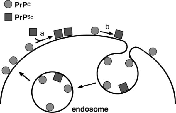
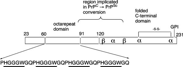
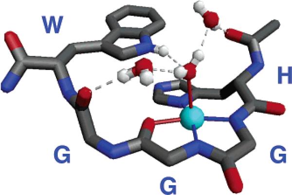
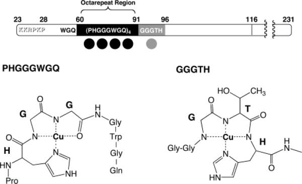
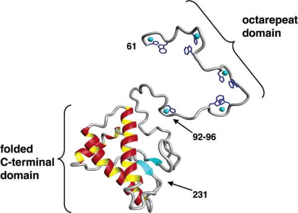
A conformational change of the prion protein is responsible for a class of neurodegenerative diseases called the transmissible spongiform encephalopathies that include mad cow disease and the human afflictions kuru and Creutzfeldt-Jakob disease. Despite the attention given to these diseases, the normal function of the prion protein in healthy tissue is unknown. Research over the past few years, however, demonstrates that the prion protein is a copper binding protein with high selectivity for Cu(2+). The structural features of the Cu(2+) binding sites have now been characterized and are providing important clues about the normal function of the prion protein and perhaps how metals or loss of protein function play a role in disease. The link between prion protein and copper may provide insight into the general, and recently appreciated, role of metals in neurodegenerative disease.


Figures

FIGURE 1
FIGURE 1
Localization and conformations of the prion protein. PrPC represents the normal form of the prion protein found in healthy tissue; PrPSc is the conformer responsible for the TSEs. PrP is tethered to cell membranes through a GPI anchor. Conversion from PrPC to PrPSc takes place either through a templating interaction with PrPSc (a) or by a spontaneous event (b). PrPC is cycled through endocytosis.
FIGURE 2
FIGURE 2
Organization of PrP and the octarepeat domain. This schematic of PrPC locates the globular C-terminal domain, the glycosylphosphatidylinositol (GPI) membrane anchor, and the octarepeat domain. Also shown is a flexible region implicated in multimerization that accompanies PrPC → PrPSc conversion. Cu2+ binding within the octarepeats involves the specific residues HGGGW (underlined).
FIGURE 3
FIGURE 3
Crystal structure of the Cu–HGGGW complex. Equatorial Cu2+ coordination is from the histidine imidazole, the deprotonated amides from the next two glycines, and the amide carbonyl of the second glycine. In addition, the NH of the indole is within hydrogen bonding distance to the oxygen of the axial water. Two additional intramolecular ordered water molecules are also shown.
FIGURE 4
FIGURE 4
Location and molecular features of the five main copper binding sites in PrP. Bond-line models of the equatorial Cu2+ coordination spheres for the two different types of binding are shown below. The structure for the single HGGGW–Cu2+ unit was determined from crystallographic and spectroscopic data. This structure is maintained for each HGGGW unit in the full octarepeat region. The coordination sphere depicted for GGGTH–Cu2+ is a model based on spectroscopic data.
FIGURE 5
FIGURE 5
Three-dimensional rendering of PrP(61–231) with coppers included. Crystal structure coordinates were used for the octarepeat binding units HGGGW. The PrP(92–96) segment was based on the model shown in Figure 4. Intervening regions were built in a relaxed conformation.

References

    1. Prusiner SB. Prion diseases and the BSE crisis. Science. 1997;278:245–251. - PubMed
    1. Prusiner SB. Prions. Proc. Natl. Acad. Sci. U.S.A. 1998;95:13363–13383. - PMC - PubMed
    1. Dobson CM. Protein misfolding, evolution and disease. Trends Biochem. Sci. 1999;24:329–332. - PubMed
    1. Caughey B, Chesebro B. Prion Protein and the Transmissible Spongiform Encephalopathies. Trends Cell Biol. 1997;7:56–62. - PubMed
    1. Sailer A, Bueler H, Fischer M, Aguzzi A, Weissmann C. No propagation of prions in mice devoid of PrP. Cell. 1994;77:967–968. - PubMed

Publication types