. 2004 Mar;5(3):304-10.
doi: 10.1038/sj.embor.7400091. Epub 2004 Feb 13.

Molecular insights into the interaction of PYM with the Mago-Y14 core of the exon junction complex

Affiliations

Molecular insights into the interaction of PYM with the Mago-Y14 core of the exon junction complex

Fulvia Bono et al. EMBO Rep. 2004 Mar.

Abstract

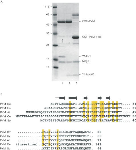
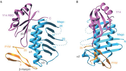
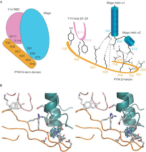
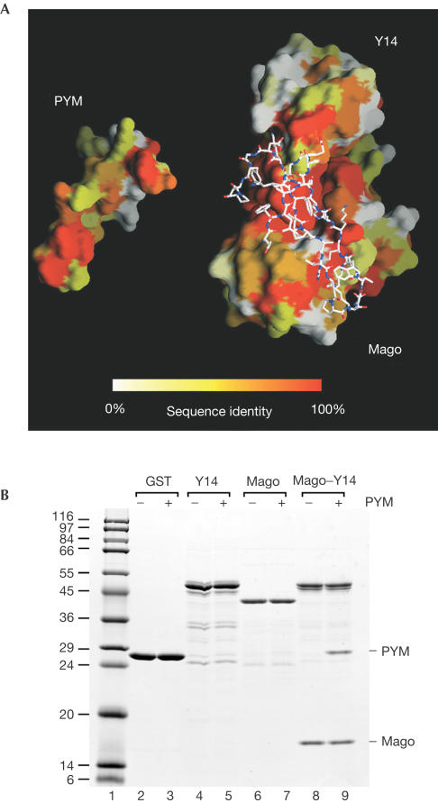
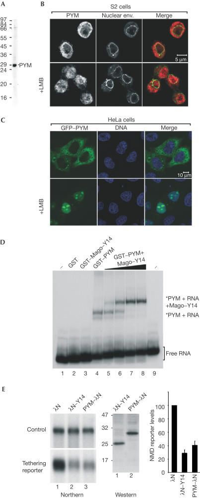
The exon junction complex (EJC) is deposited on mRNAs as a consequence of splicing and influences postsplicing mRNA metabolism. The Mago-Y14 heterodimer is a core component of the EJC. Recently, the protein PYM has been identified as an interacting partner of Mago-Y14. Here we show that PYM is a cytoplasmic RNA-binding protein that is excluded from the nucleus by Crm1. PYM interacts directly with Mago-Y14 by means of its N-terminal domain. The crystal structure of the Drosophila ternary complex at 1.9 A resolution reveals that PYM binds Mago and Y14 simultaneously, capping their heterodimerization interface at conserved surface residues. Formation of this ternary complex is also observed with the human proteins. Mago residues involved in the interaction with PYM have been implicated in nonsense-mediated mRNA decay (NMD). Consistently, human PYM is active in NMD tethering assays. Together, these data suggest a role for PYM in NMD.


Figures

Figure 1
Figure 1
The N-terminal domain of PYM is sufficient to interact with Mago–Y14. (A) Pull-down experiments in which lysates from E. coli expressing the D. melanogaster proteins indicated on the right were incubated with glutathione agarose beads. The construct Y14ΔC includes residues 1–154 and Y14ΔNΔC includes residues 67–154. (B) Sequence alignment of PYM N-terminal region from D. melanogaster (Dm), H. sapiens (Hs), Arabidopsis thaliana (At), C. elegans (Ce) and S. pombe (Sp). Secondary structure elements are shown above the sequences. Conserved residues are highlighted in yellow. The presence of an insertion in the Ce orthologue is indicated.
Figure 2
Figure 2
Structure of the Drosophila PYM–Mago–Y14 ternary complex. (A) View of the complex between the RBD domain of Y14 (pink), Mago (cyan) and the N-terminal domain of PYM (orange). PYM binds at the edge of the Y14 β-sheet (β2–β3 loop) and at the edge of the Mago α-helices, spanning both components of the heterodimer. Dotted lines represent the approximate path of loops in Mago that were disordered and not modelled. (B) View of the complex after a 90° rotation about the vertical axis with respect to the view in (A).
Figure 3
Figure 3
PYM binds Mago–Y14 with extensive interactions. (A) Schematic view of the PYM–Mago–Y14 complex (left panel) and schematic diagram highlighting the key residues involved in the interaction (right panel). Positively charged residues of PYM interact with negatively charged residues of Mago helices α1 and α2. In addition, PYM interacts with the β2–β3 loop of Y14 by means of hydrophobic contacts. Colours are as in Fig 2. Hydrogen bonds are shown with dotted lines. (B) Stereo representation of the structure and of the interacting residues in a similar orientation as in Figs 2A,3A.
Figure 4
Figure 4
The PYM–Mago–Y14 complex is conserved across species. (A) The interaction surfaces of D. melanogaster PYM and the Mago–Y14 heterodimer have been opened up relative to the view in Fig 2A. The two surfaces are coloured according to sequence conservation, ranging from orange for conserved residues to white for variable residues. On the right, the atomic model of PYM is shown bound to the surface of Mago–Y14. (B) Lysates prepared from E. coli expressing untagged H. sapiens (Hs) PYM were incubated with glutathione agarose beads coated with GST, GST–Hs Y14, GST–Hs Mago or GST–Hs Y14–Mago dimers.
Figure 5
Figure 5
PYM is a cytosolic RNA-binding protein involved in NMD. (A) Western blot with purified anti-Dm PYM antibody showing that a unique band of the right size is recognized in S2 cell extracts. (B) Immunolocalization of endogenous PYM in S2 cells. Endogenous PYM (red) is detected predominantly in the cytoplasm in untreated S2 cells, and in both the nucleoplasm and cytoplasm in cells treated for 12 h with leptomycin B (LMB). The nuclear envelope is stained with fluorescently labelled wheat germ agglutinin (WGA, green). (C) Subcellular localization of human PYM. HeLa cells were transfected with a plasmid expressing GFP–PYM. The fusion protein (green) is detected throughout the cytoplasm and excluded from the nucleoplasm. Following LMB treatment (3 h), GFP–PYM accumulates within the nucleus and the nucleolus. DNA is stained with Hoechst (blue). (D) Gel-mobility assay performed with a labelled RNA probe and the purified recombinant proteins indicated above the lanes. Dm Mago and PYM were N-terminally fused to GST. In lanes 2–4, the concentration of the recombinant proteins was 0.1 μg/μl. Lanes 5–8 show binding reactions with 0.1 μg/μl of GST–PYM supplemented with 0.005, 0.02, 0.05 and 0.1 μg/μl of GST Dm Mago–Y14 dimers, respectively. The position of the free RNA probe (lanes 1 and 9) and of the RNA-bound complexes is shown on the right. (E) NMD assay. Tethering PYM–λN to the 3′UTR of a reporter mRNA results in degradation of the reporter to a similar extent as observed when λN–Y14 is tethered. The steady-state levels of the NMD reporter in the presence of PYM or Y14 were quantified in three independent experiments, normalized to those of the transfection control and expressed as a percentage of the levels observed when the λN-peptide alone was coexpressed. The expression levels of the proteins were analysed by western blot with an anti-λN antibody.

References

    1. Brünger AT et al. ( 1998) Crystallographic and NMR system: a new software system for macromolecular structure determination. Acta Crystallogr D 54: 905–921 - PubMed
    1. CCP4 ( 1994) The CCP4 suite: programs for protein crystallography. Acta Crystallogr D 50: 760–763 - PubMed
    1. Forler D, Kocher T, Rode M, Gentzel M, Izaurralde E, Wilm M ( 2003) An efficient protein complex purification method for functional proteomics in higher eukaryotes. Nat Biotech 21: 89–92 - PubMed
    1. Forler D, Rabut G, Ciccarelli FD, Herold A, Kocher T, Niggeweg R, Bork P, Ellenberg J, Izaurralde E ( 2004) RanBP2/Nup358 provides a major binding site for NXF1-p15 dimers at the nuclear pore complex and functions in nuclear mRMA export. Mol Cell Biol 24: 1155–1167 - PMC - PubMed
    1. Fribourg S, Gatfield D, Izaurralde E, Conti E ( 2003) A novel mode of RBD-protein recognition in the Y14–Mago complex. Nat Struct Biol 10: 433–439 - PubMed

Publication types

MeSH terms

Associated data