

Glial tumor grading and outcome prediction using dynamic spin-echo MR susceptibility mapping compared with conventional contrast-enhanced MR: confounding effect of elevated rCBV of oligodendrogliomas [corrected]
- PMID: 14970020
- PMCID: PMC7974605
Glial tumor grading and outcome prediction using dynamic spin-echo MR susceptibility mapping compared with conventional contrast-enhanced MR: confounding effect of elevated rCBV of oligodendrogliomas [corrected]
Erratum in
- AJNR Am J Neuroradiol. 2004 Mar;25(3):B1
Abstract
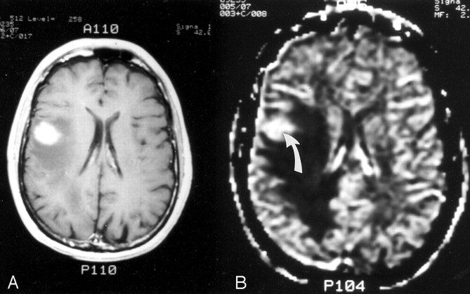
Background and purpose: The MR imaging characteristics of oligodendrogliomas and astrocytomas on spin-echo (SE), echo-planar relative cerebral blood volume (rCBV) maps, to our knowledge, have not previously been emphasized. We compared the specificity of SE rCBV mapping with that of conventional, contrast material-enhanced MR imaging in differentiating high- from low-grade glial tumors and in predicting survival of patients with these lesions.
Methods: Thirty consecutive adult patients with suspected gliomas underwent conventional and rCBV MR imaging. Representative maximal rCBV regions of interest were chosen from each lesion. Resultant values were normalized to those of corresponding, contralateral, uninvolved regions. These normalized CBV (nCBV) values were correlated with degree of contrast enhancement, histopathologic tumor grade, and survival.
Results: Twenty-two patients had astroctyomas and eight had oligodendrogliomas. With an nCBV cutoff ratio of 1.5, 13 of 13 high-grade astrocytomas were correctly categorized, three of which did not enhance. Seven of nine low-grade astrocytomas were correctly classified by their nCBV values, including one enhancing lesion. Of eight oligodendrogliomas, four of four high-grade and two of four low-grade tumors had elevated nCBV values; two low-grade oligodendrogliomas enhanced, one with nCBV greater than 1.5 and one with nCBV less than 1.5. In 19 patients with astrocytoma for whom survival data were available, correlation with survival was better for nCBV (mean survival 91 +/- 14 months for nCBV < 1.5 versus 24 +/- 27 months for nCBV > 1.5, P <.0001) than for enhancement (mean survival 61 +/- 35 months without enhancement versus 22 +/- 29 months with enhancement, P =.03).
Conclusion: Elevated SE rCBV was a sensitive, but not specific, marker for high-grade histopathology: all high-grade tumors had nCBV foci values greater than 1.5. No tumor with nCBV region of interest less than 1.5 was high grade (100% predictive value for excluding high grade). Degree of nCBV elevation was a stronger predictor of both tumor grade and survival than was degree of enhancement. A significant proportion of low-grade glial neoplasms, most notably oligodendrogliomas, may display high rCBV foci not reflective of high-grade histopathology.
Figures


References
-
- Kleihues P, Burger PC, Scheithauer BW. Histological Typing of Tumors of the Central Nervous System, 2nd ed. Berlin: Springer-Verlag;1993
-
- Dumas-Duport C, Scheithauer B, O’Fallon J, et al. Grading of astrocytomas: a simple and reproducible method. Cancer 1988;62:2152–2165 - PubMed
-
- Scatliff JH, Radcliffe WB, Pittman HH, Park CH. Vascular structure of glioblastomas. Radium Ther Nucl Med 1969;105:795–805 - PubMed
-
- Brem S, Cotran R, Folkman J. Tumor angiogenesis: a quantitative method for histologic grading. J Natl Cancer Inst 1972;48:347–356 - PubMed
-
- Leon SP, Folkerth RD, Black PM. Microvessel density is a prognostic indicator for patients with astroglial brain tumors. Cancer 1996;77:362–372 - PubMed
Publication types
MeSH terms
Grants and funding
LinkOut - more resources
Full Text Sources
Medical