Paramagnetic effect of supplemental oxygen on CSF hyperintensity on fluid-attenuated inversion recovery MR images
- PMID: 14970030
- PMCID: PMC7974613
Paramagnetic effect of supplemental oxygen on CSF hyperintensity on fluid-attenuated inversion recovery MR images
Abstract
Background and purpose: Oxygen has a known paramagnetic effect and increases CSF signal intensity on fluid-attenuated inversion recovery (FLAIR) MR images. The purposes of this study were to investigate the effect of supplemental oxygen on CSF signal intensity and the arterial partial pressure of oxygen and to determine the possible synergistic effect of oxygen and albumin on T1 shortening effect in vitro.
Methods: Six healthy volunteers underwent FLAIR MR imaging of the brain before and during inhalation of 10 to 15 L/min of 100% oxygen for < or = 30 min. The signal intensity was measured in the subarachnoid spaces and various tissues and correlated with estimated arterial partial pressure of oxygen and arterial carbon dioxide pressure. In vitro measurements were also obtained by using two sets of saline-filled tubes with various concentrations of albumin, one of which was exposed to increased oxygen levels. In vitro T1 relaxation times were calculated to assess the possible synergistic effect of oxygen and albumin.
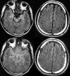
Results: FLAIR images of healthy volunteers showed increased CSF signal intensity within the basal cisterns and sulci along the cerebral convexities. The CSF hyperintensity was observed immediately after the initiation of supplemental oxygen and remained stable during the oxygen administration. There was approximately a 4- to 5.3-fold increase in signal intensity with supplemental oxygen. The phantom experiments showed a T1 shortening effect of oxygen. Albumin significantly altered T1 relaxation time only at high concentrations of albumin.
Conclusion: Inhalation of increased levels of oxygen led to readily detectable CSF hyperintensity on FLAIR images of healthy volunteers. No significant synergetic effect of albumin and oxygen was noted.
Figures


Similar articles
-
Supplemental oxygen causes increased signal intensity in subarachnoid cerebrospinal fluid on brain FLAIR MR images obtained in children during general anesthesia.Radiology. 2004 Oct;233(1):51-5. doi: 10.1148/radiol.2331031375. Radiology. 2004. PMID: 15454616 Clinical Trial.
-
Cerebrospinal fluid signal intensity increase on FLAIR MR images in patients under general anesthesia: the role of supplemental O2.Radiology. 2001 Jan;218(1):152-6. doi: 10.1148/radiology.218.1.r01ja43152. Radiology. 2001. PMID: 11152794
-
Fraction of inspired oxygen in relation to cerebrospinal fluid hyperintensity on FLAIR MR imaging of the brain in children and young adults undergoing anesthesia.AJR Am J Roentgenol. 2002 Sep;179(3):791-6. doi: 10.2214/ajr.179.3.1790791. AJR Am J Roentgenol. 2002. PMID: 12185066
-
Abnormal hyperintensity within the subarachnoid space evaluated by fluid-attenuated inversion-recovery MR imaging: a spectrum of central nervous system diseases.Eur Radiol. 2003 Dec;13 Suppl 4:L192-201. doi: 10.1007/s00330-003-1877-9. Eur Radiol. 2003. PMID: 15018187 Review.
-
Hyperintensity in the subarachnoid space on FLAIR MRI.AJR Am J Roentgenol. 2007 Oct;189(4):913-21. doi: 10.2214/AJR.07.2424. AJR Am J Roentgenol. 2007. PMID: 17885065 Review.
Cited by
-
Effects of superficial temporal artery to middle cerebral artery (STA-MCA) anastomosis on the cerebrospinal fluid gas tensions and pH in hemodynamically compromised patients.Acta Neurochir (Wien). 2023 Dec;165(12):3709-3715. doi: 10.1007/s00701-023-05855-5. Epub 2023 Oct 26. Acta Neurochir (Wien). 2023. PMID: 37882875
-
Reduction of Oxygen-Induced CSF Hyperintensity on FLAIR MR Images in Sedated Children: Usefulness of Magnetization-Prepared FLAIR Imaging.AJNR Am J Neuroradiol. 2016 Aug;37(8):1549-55. doi: 10.3174/ajnr.A4723. Epub 2016 Mar 17. AJNR Am J Neuroradiol. 2016. PMID: 26988816 Free PMC article.
-
Intraoperative monitoring of cerebrospinal fluid gas tension and pH before and after surgical revascularization for moyamoya disease.Surg Neurol Int. 2024 May 10;15:158. doi: 10.25259/SNI_281_2024. eCollection 2024. Surg Neurol Int. 2024. PMID: 38840605 Free PMC article.
-
Proper fraction of inspired oxygen for reduction of oxygen-induced canine cerebrospinal fluid hyperintensity on fluid attenuation inversion recovery sequence using low-field magnetic resonance imaging.J Vet Med Sci. 2020 Oct 7;82(9):1321-1328. doi: 10.1292/jvms.20-0060. Epub 2020 Jul 20. J Vet Med Sci. 2020. PMID: 32684615 Free PMC article.
-
Use of diffusion tensor imaging to assess the impact of normobaric hyperoxia within at-risk pericontusional tissue after traumatic brain injury.J Cereb Blood Flow Metab. 2014 Oct;34(10):1622-7. doi: 10.1038/jcbfm.2014.123. Epub 2014 Jul 9. J Cereb Blood Flow Metab. 2014. PMID: 25005875 Free PMC article.
References
-
- Oatridge A, Hajnal JV, Cowan FM, Baudouin CJ, Young IR, Bydder GM. MRI diffusion-weighted imaging of the brain: contributions to image contrast from CSF signal reduction, use of a long echo time and diffusion effects. Clin Radiol 1993;47:82–90 - PubMed
-
- Singer MB, Atlas SW, Drayer BP. Subarachnoid space disease: diagnosis with fluid-attenuated inversion-recovery MR imaging and comparison with gadolinium-enhanced spin-echo MR imaging–blinded reader study. Radiology 1998;208:417–422 - PubMed
MeSH terms
Substances
LinkOut - more resources
Full Text Sources
Other Literature Sources
Medical
