

Noninvasive MR cisternography with fluid-attenuated inversion recovery and 100% supplemental O(2) in the evaluation of neurocysticercosis
Affiliations
- PMID: 14970035
- PMCID: PMC7974615
Item in Clipboard
Noninvasive MR cisternography with fluid-attenuated inversion recovery and 100% supplemental O(2) in the evaluation of neurocysticercosis
AJNR Am J Neuroradiol.
2004 Feb.
Erratum in
- AJNR Am J Neuroradiol. 2004 Mar;25(3):B1
Abstract
We describe an MR protocol for the noninvasive imaging of the subarachnoid space, which we use in patients with suspected neurocysticercosis in this space. It consists of a fluid-attenuated inversion recovery sequence performed 5 minutes after the continuous inhalation of 100% O(2) with a resultant increase in the signal intensity of the CSF that leads to a greater conspicuity of cyst walls in relation to the cortex and the extraventricular CSF.
Figures
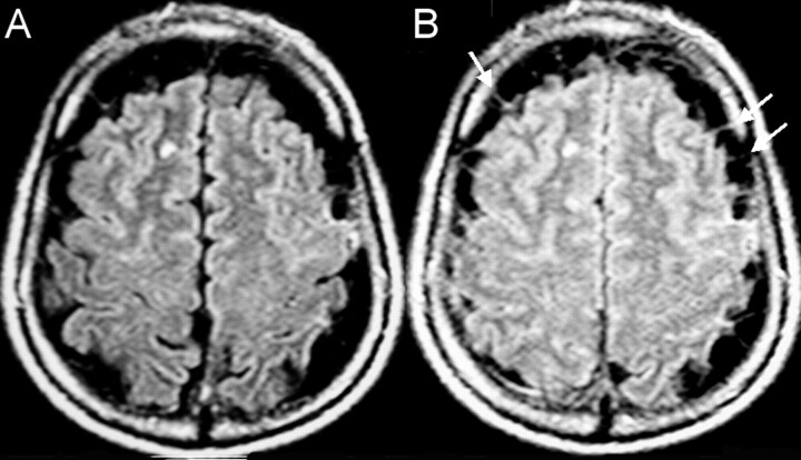
A 40-year-old man with parenchymal neurocysticercosis and suspected subarachnoid involvement. A, MR FLAIR image (1.0 T; TR, 11,000 ms; TE, 140 ms; TI, 2600 ms) shows no extraaxial lesions. B, FLAIR image after 100% O2 for 5 minutes shows two small cysts in the Sylvian fissures confirming racemous neurocysticercosis.
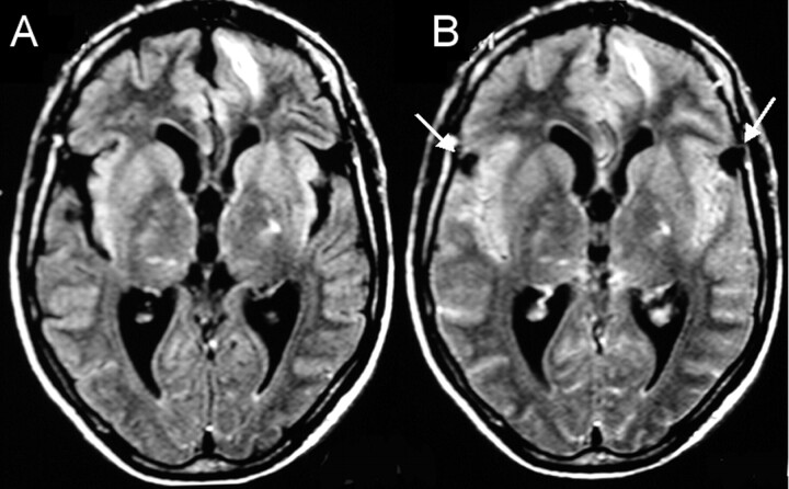
A 57-year-old man with multiple vesicular lesions in the brain parenchyma. A, FLAIR image shows prominent CSF space along the cerebral convexities. B, FLAIR image after 100% O2 for 5 minutes does not show the expected increased signal intensity of the CSF due to the presence of multiple clustered extraaxial cysts. The walls of the cysts (arrows) are better visualized after 100% O2.
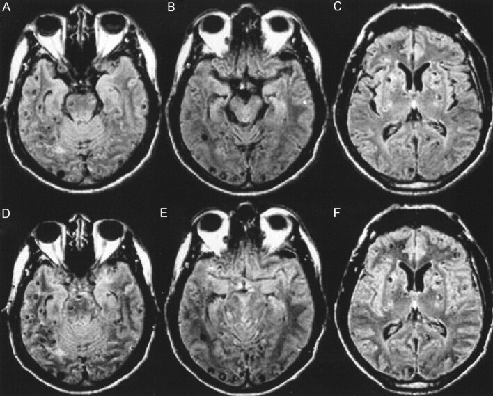
A 34-year-old male patient with known neurocysticercosis. A, B, and C, FLAIR images show multiple vesicular lesions in the parenchyma, mainly along the cortical surfaces. The perimesencephalic cisterns are prominent and contain hyperintense foci that could represent the expected flow artifact or cystic lesions with scoleces (B). D, E, and F, FLAIR after 100% O2 shows increased signal intensity in the sulci and basal cisterns, allowing the visualization of a greater number of cortical lesions in the frontal, temporal, and occipital lobes. Also, the increased signal intensity of the CSF confirms that there is no cystic lesion in the perimesencephalic cisterns (E).
Similar articles
-
Paramagnetic effect of supplemental oxygen on CSF hyperintensity on fluid-attenuated inversion recovery MR images.AJNR Am J Neuroradiol. 2004 Feb;25(2):274-9. AJNR Am J Neuroradiol. 2004. PMID: 14970030 Free PMC article.
-
Relevance of 3D magnetic resonance imaging sequences in diagnosing basal subarachnoid neurocysticercosis.Acta Trop. 2015 Dec;152:60-65. doi: 10.1016/j.actatropica.2015.08.017. Epub 2015 Aug 29. Acta Trop. 2015. PMID: 26327445
-
Supplemental oxygen causes increased signal intensity in subarachnoid cerebrospinal fluid on brain FLAIR MR images obtained in children during general anesthesia.Radiology. 2004 Oct;233(1):51-5. doi: 10.1148/radiol.2331031375. Radiology. 2004. PMID: 15454616 Clinical Trial.
-
Neurocysticercosis: evaluation with advanced magnetic resonance techniques and atypical forms.Top Magn Reson Imaging. 2005 Apr;16(2):127-44. doi: 10.1097/01.rmr.0000189106.78146.98. Top Magn Reson Imaging. 2005. PMID: 16340333 Review.
-
CT and MR imaging of neurocysticercosis.AJR Am J Roentgenol. 1999 Dec;173(6):1485-90. doi: 10.2214/ajr.173.6.10584787. AJR Am J Roentgenol. 1999. PMID: 10584787 Review. No abstract available.
Cited by
-
Hydrocephalus in neurocysticercosis.Childs Nerv Syst. 2011 Oct;27(10):1709-21. doi: 10.1007/s00381-011-1500-3. Epub 2011 Sep 17. Childs Nerv Syst. 2011. PMID: 21928035 Review.
-
Cysticercosis: Recent Advances in Diagnosis and Management of Neurocysticercosis.Curr Infect Dis Rep. 2005 Jan;7(1):39-47. doi: 10.1007/s11908-005-0022-0. Curr Infect Dis Rep. 2005. PMID: 15610670
-
Dynamic oxygen-enhanced MRI of cerebrospinal fluid.PLoS One. 2014 Jun 23;9(6):e100723. doi: 10.1371/journal.pone.0100723. eCollection 2014. PLoS One. 2014. PMID: 24956198 Free PMC article.
-
Imaging of cerebellopontine angle lesions: an update. Part 2: intra-axial lesions, skull base lesions that may invade the CPA region, and non-enhancing extra-axial lesions.Eur Radiol. 2007 Nov;17(11):2908-20. doi: 10.1007/s00330-007-0680-4. Epub 2007 Jun 14. Eur Radiol. 2007. PMID: 17569053 Review.
-
Neuroimaging in Central Nervous System Infections.Curr Neurol Neurosci Rep. 2017 Jun;17(6):49. doi: 10.1007/s11910-017-0756-8. Curr Neurol Neurosci Rep. 2017. PMID: 28466277 Review.
References
-
- Zeng Q, Xiong L, Jinkins JR, et al. Intrathecal gadolinium-enhanced MR myelography and cisternography: a pilot study in human patients. AJR Am J Roentgenol 1999;173:1109–1115 - PubMed
-
- Jinkins JR, Rudwan M, Krumina G, Tali ET. Intrathecal gadolinium-enhanced MR cisternography in the evaluation of clinically suspected cerebrospinal fluid rhinorrhea in humans: early experience. Radiology 2002;222:555–559 - PubMed
-
- Tali ET, Ercan N, Krumina G, et al. Intrathecal gadolinium (gadopentetate dimeglumine) enhanced magnetic resonance myelography and cisternography: results of a multicenter study. Invest Radiol 2002;37:152–159 - PubMed
-
- Deliganis AV, Fisher DJ, Lam AM, Maravilla KR. Cerebrospinal fluid signal intensity increase on FLAIR MR images in patients under general anesthesia: the role of supplemental O2. Radiology 2001;218:152–156 - PubMed
MeSH terms
Substances
LinkOut - more resources
Full Text Sources
Medical