Comparative Study
. 2004 Feb;25(2):298-306.

Endovascular treatment of intracranial wide-necked aneurysms using three-dimensional coils: predictors of immediate anatomic and clinical results

Affiliations
Comparative Study

Endovascular treatment of intracranial wide-necked aneurysms using three-dimensional coils: predictors of immediate anatomic and clinical results

Jean-Noël Vallée et al. AJNR Am J Neuroradiol. 2004 Feb.

Abstract

Background and purpose: Aneurysms with a wide neck constitute a persistent challenge for endovascular therapy with coils. Our purpose was to evaluate the immediate anatomic and clinical results of treating intracranial wide-necked aneurysms by using three-dimensional (3D) coils.

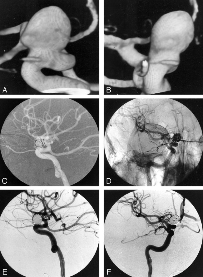
Methods: During a 2-year period, 160 aneurysms (116 with a neck < or = 4 mm, group A; 44 with a neck > 4 mm, group B) in 157 patients in eight participating centers were consecutively treated. The procedure consisted first of framing the aneurysm with one or more 3D spherical coils and then filling it with helical coils. Results were evaluated with univariate analysis. Multivariate analysis was used to identify independent predictors of these results.

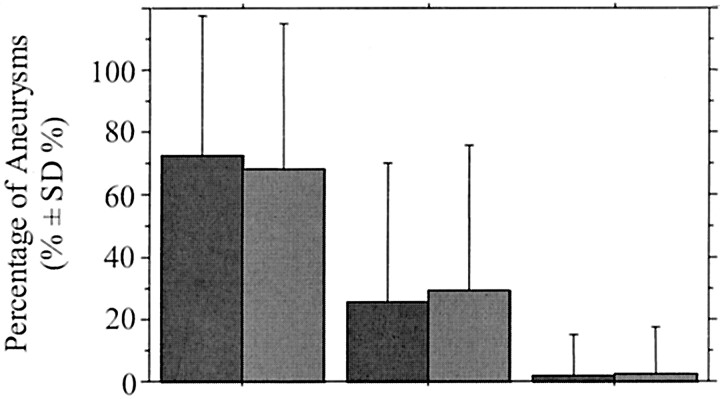
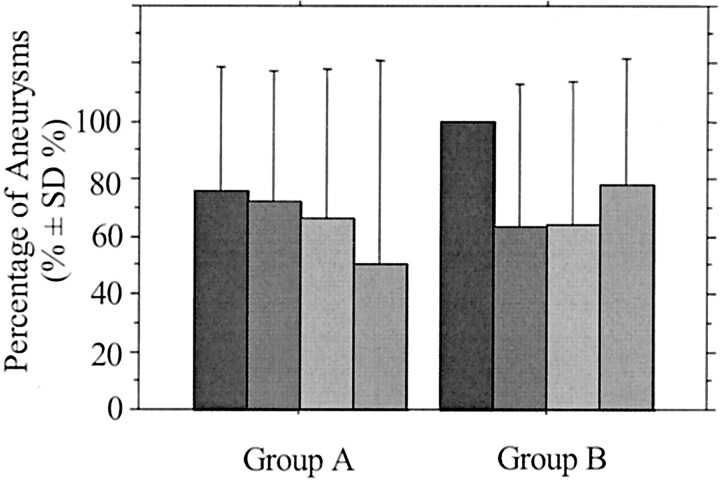
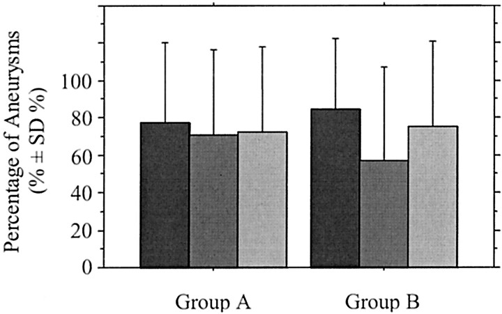
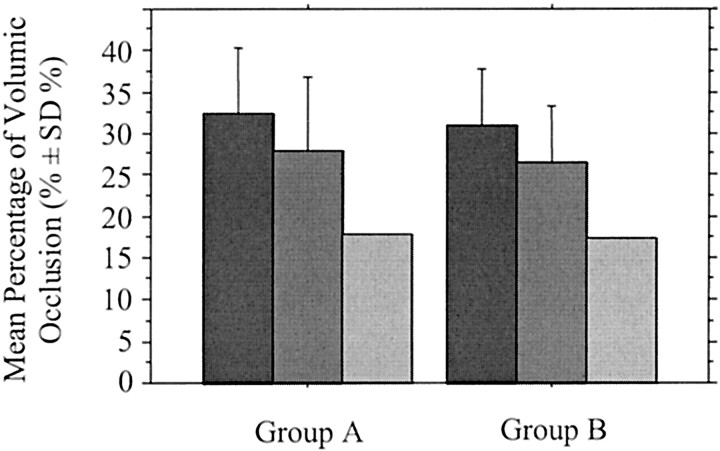
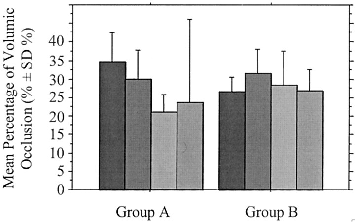
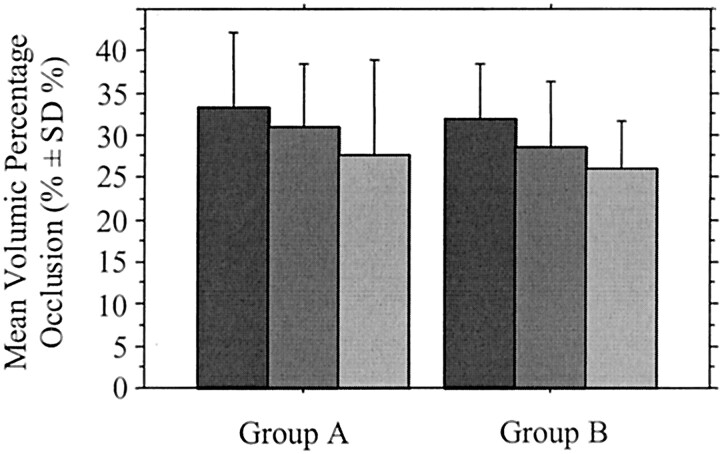
Results: Angiographic occlusion was complete in 84 (72%) and 30 (68%) aneurysms in groups A and B, respectively. Mean percentage of volumic occlusion in these groups was 30.9% and 29.2%, respectively. Perioperative morbidity and mortality rates were 4%, respectively, in group A and 2%, respectively, in group B. No significant difference between the two groups was observed. However, percentage of volumic occlusion correlated with sac-to-neck ratio less than 1.5 (P =.061) and with sac size (P =.002) except when three or more 3D coils per aneurysm were used (P =.222). The better the percentage of volumic occlusion, the better the degree of angiographic occlusion (P =.004). Percentage of volumic occlusion was an independent predictor of angiographic complete occlusion (P =.001). World Federation of Neurological Surgeons subarachnoid hemorrhage scale grade 5 was an independent predictor of perioperative mortality (P =.043).

Conclusion: Three-dimensional coils proved to be useful for improving coil packing and angiographic and volumic occlusion of aneurysms with a neck greater than 4 mm, at the time of treatment, provided the sac-to-neck ratio was 1.5 or greater, and the largest number of 3D coils were first positioned.


Figures

F<sc>ig</sc> 1.
Fig 1.
Pre- and posttreatment images of an unruptured intracranial aneurysm in a 46-year-old woman who underwent occlusion with 3D Micrus coils. A and B, Lateral (A) and anteroposterior (B) 3D angiograms of the right internal carotid artery show a periophthalmic aneurysm with a maximum sac size of 9 mm and a neck size of 6 mm. C and D, Lateral road mapping image (C) and oblique unsubtracted angiogram (D) of the right internal carotid artery after placement of the first spherical coil (9-mm coil loop diameter) show that the 3D configuration of the coil provides an anatomically compliant frame within the aneurysm and a scaffold that covers the neck. E and F, Lateral (E) and anteroposterior (F) subtracted posttreatment angiograms show complete occlusion of the aneurysm.
F<sc>ig</sc> 2.
Fig 2.
Graph shows percentage of aneurysms according to degree of angiographic aneurysmal occlusion at completion of the initial endovascular treatment, in group A ([dark gray bars] aneurysms with a neck 4 mm) and group B ([medium gray bars] aneurysms with a neck >4 mm). Degree of angiographic occlusion was not significantly different between the two groups (P = .696).
F<sc>ig</sc> 3.
Fig 3.
Graph shows percentage of completely occluded aneurysms according to sac size at completion of the initial endovascular treatment in group A (neck ≤ 4 mm) and group B (neck >4 mm). Black bars indicate sac size <5 mm; dark gray bars, sac size 5–10 mm; medium gray bars, sac size 10–15 mm; light gray bars, sac size 15–25 mm.
F<sc>ig</sc> 4.
Fig 4.
Graph shows percentage of completely occluded aneurysms according to sac-to-neck size ratio at completion of the initial endovascular treatment in group A (neck ≤ 4 mm) and group B (neck >4 mm). Dark gray bars indicate ratio ≥3; medium gray bars, ratio 1.5–3; light gray bars, ratio <1.5.
F<sc>ig</sc> 5.
Fig 5.
Graph shows the mean percentage of volumic aneurysmal occlusion according to degree of angiographic occlusion at completion of the initial endovascular treatment in group A (neck 4 mm) and group B (neck >4 mm). Percentage of volumic occlusion was not significantly different between the two groups (P = .247). Dark gray bars indicate complete occlusion; medium gray bars, near complete occlusion; light gray bars, incomplete occlusion.
F<sc>ig</sc> 6.
Fig 6.
Graph shows the mean percentage of volumic aneurysmal occlusion according to sac size at completion of the initial endovascular treatment in group A (neck 4 mm) and group B (neck >4 mm). Black bars indicate sac size <5 mm; dark gray bars, sac size 5–10 mm; medium gray bars, sac size 10–15 mm; light gray bars, sac size 15–20 mm.
F<sc>ig</sc> 7.
Fig 7.
Graph shows the mean percentage of volumic aneurysmal occlusion according to the sac-to-neck size ratio at completion of the initial endovascular treatment in group A (neck 4 mm) and group B (neck >4 mm). Dark gray bars indicate ratio ≥3; medium gray bars, ratio 1.5–3; light gray bars, ratio <1.5.

Similar articles

Cited by

References

    1. Guglielmi G, Viñuela F, Dion J, Duckwiler G. Electrothrombosis of saccular aneurysms via endovascular approach, II: preliminary clinical experience. J Neurosurg 1991;75:8–14 - PubMed
    1. Horowitz MB, Levy E, Kassam A, Purdy PD. Endovascular therapy for intracranial aneurysms: a historical and present status review. Surg Neurol 2002;57:147–158 - PubMed
    1. Zubillaga AF, Guglielmi G, Viñuela F, et al. Endovascular occlusion of intracranial aneurysms with electrically detachable coils: correlation of aneurysm neck size and treatment results. AJNR Am J Neuroradiol 1994;15:815–820 - PMC - PubMed
    1. Viñuela F, Duckwiler G, Mawad M. Guglielmi detachable coil embolization of acute intracranial aneurysm: perioperative anatomical and clinical outcome in 403 patients. J Neurosurg 1997;86:475–482 - PubMed
    1. Ayton Hope JK, Byrne JV, Molyneux AJ. Factors influencing successful angiographic occlusion of aneurysms treated by coil embolization. AJNR Am J Neuroradiol 1999;20:391–399 - PMC - PubMed

MeSH terms