

Case Reports
External jugular vein vascular malformation: sonographic and MR imaging appearances
Affiliations
- PMID: 14970043
- PMCID: PMC7974618
Item in Clipboard
Case Reports
External jugular vein vascular malformation: sonographic and MR imaging appearances
AJNR Am J Neuroradiol.
2004 Feb.
Abstract
Vascular malformations arising from the wall of the external jugular vein are rare. This case series discusses the sonographic and MR imaging appearances of four such cases and reviews the literature. The diagnosis should be suggested preoperatively particularly because of the close relationship such malformations to the external jugular vein, as this helps surgeons to plan the operative procedure. The imaging appearances are similar to those of other vascular malformations elsewhere in the head and neck.
Figures
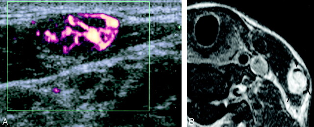
Case 1. Sonographic and MR images obtained in a 31-year-old Indonesian woman with a vascular malformation of the left external jugular vein. A, Transverse power Doppler sonography shows vascularity within a well-defined, hypoechoic, heterogeneous mass. B, Axial T1-weighed fat-suppressed contrast-enhanced MR imaging image (TR/TE, 450/15) shows intense enhancement of the mass (arrows) and its relationship to the left external jugular vein (arrowhead).
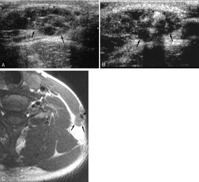
Case 2. Sonographic and MR images obtained in a 21-year-old woman with a left external jugular vein vascular malformation. A, Transverse gray-scale sonography shows a hypoechoic heterogeneous mass (arrows) closely related to the external jugular vein (arrowheads). B, Longitudinal gray-scale sonography shows a phlebolith (arrowheads) within the mass (arrows). C, Axial T1-weighed spin-echo MR imaging image (TR/TE, 425/12) shows an isointense mass (arrows) closely related to the left external jugular vein (arrowhead). It is closely abutting the posterior edge of the left sternocleidomastoid muscle (asterisk).
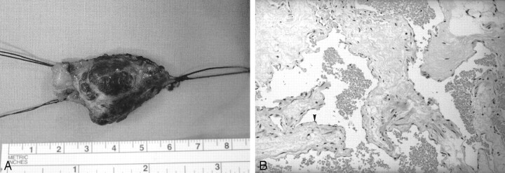
Case 2. Pathologic and histologic sections of a specimen obtained by excision of the left external jugular vein vascular malformation. A, Gross specimen of the excised lesion shows a well-circumscribed mass with the proximal and distal vascular stumps of the external jugular vein. B, High-power histologic view of the vascular channels. Note the lining flattened endothelial cells (arrowhead) and the red cells inside the lumens. (Hematoxylin-eosin stain, original magnification, ×120.)
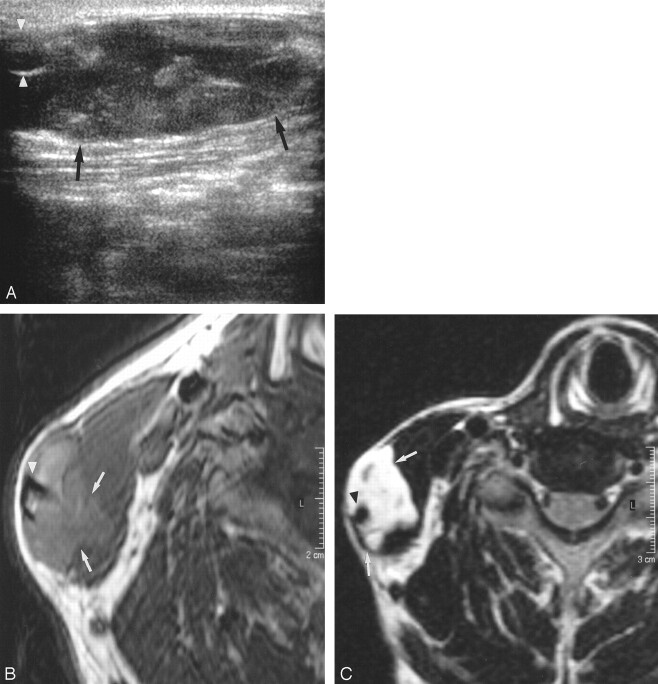
Case 3. Sonographic and MR images obtained in a 37-year-old man with right external jugular vein vascular malformation. A, Transverse gray-scale sonography shows an ill-defined, hypoechoic, heterogeneous mass (arrows) inseparable from the external jugular vein (arrowheads). B, Axial T1-weighed spin-echo MR imaging image (TR/TE, 425/18) shows a slightly hyperintense mass (arrows) with ill-defined edges closely related to the right external jugular vein (arrowhead). C, Axial fat-suppressed T2-weighed MR imaging image (TR/TE, 2500/108) shows a hyperintense mass (arrows) closely related to and inseparable from the right external jugular vein (arrowhead). Note its infiltration into the adjacent sternocleidomastoid muscle.
References
-
- Mulliken JB, Glowacki J Hemangiomas and vascular malformations in infants and children: a classification based on endothelial characteristics. Plast Reconstr Surg 1982;69:412–422 - PubMed
-
- Meyers MA. Hemangioma of the external jugular vein. Radiology 1967;89:483–485 - PubMed
-
- Sarteschi LM, Bonanomi G, Mosca F, Ferrari M. External jugular vein hemangioma occuring as a lateral neck mass. J Ultrasound Med 1999;18:719–721 - PubMed
-
- Gold L, Nazarian LN, Johar AS, Rao VM. Characterization of maxillofacial soft tissue vascular anomalies by ultrasound and color Doppler imaging: an adjuvant to computed tomography and magnetic resonance imaging. J Oral Maxillofac Surg 2003;61:19–31 - PubMed
-
- Yang WT, Ahuja AT, Metreweli C. Sonographic features of head and neck hemangiomas and vascular malformations: review of 23 patients. J Ultrasound Med 1997;16:39–44 - PubMed
Publication types
MeSH terms
LinkOut - more resources
Full Text Sources
Medical