

Early prenatal MR imaging diagnosis of polymicrogyria
Affiliations
- PMID: 14970044
- PMCID: PMC7974610
Item in Clipboard
Early prenatal MR imaging diagnosis of polymicrogyria
AJNR Am J Neuroradiol.
2004 Feb.
Abstract
The case of a 24-week-old fetus that showed features suggestive of focal cortical developmental anomaly at prenatal MR imaging is presented. The anomaly was confirmed to be polymicrogyria by 34-week prenatal and the 3-day postnatal MR imaging studies. The report demonstrates that the development of polymicrogyria can be assessed throughout different stages by prenatal MR imaging. In the case reported, the additional presence of periventricular heterotopia strongly suggests that a neuronal migration alteration coexisted with a postmigrational disorder.
Figures
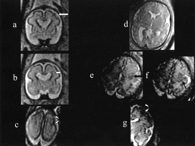
Multiplanar T2-weighted single-shot fast spin-echo sections from 24-week (A–C) and 34-week (D–G) gestational age. A, Coronal section showing absent septum pellucidum and focal irregular wartlike profile of the cortical rim in the left hemisphere (white arrow). B, Contiguous coronal section depicting a small hypointense subependymal nodule associated with a thin hypointense line extending toward the cortex (white arrowhead). C, Axial section showing an unclear area of cortical-rim abnormal profile with apparent irregular infolding in the left hemisphere (curved white arrow). D, Axial section depicting the absence of septum pellucidum. E (left) and F (right), Axial contiguous sections from the same study showing in the left posterior frontal lobe a focal area of abnormal cortical-rim profile with some irregular infolding (black arrow). G, Coronal section from the same study depicting in the left frontal lobe the same area of irregular and abnormally tight cortical gyri infolding (curved white arrow). Only the left half of the section has been reproduced, to eliminate the dynamic windowing problems related to the high signal intensity in the right side of the image, caused by the proximity to the surface coil.
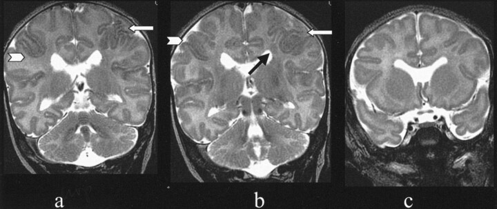
Three T2-weighted coronal FSE sections from the postnatal MR imaging study. A focal area of abnormally tight cortical infolding at the left frontal lobe cortex level is visible (white arrows). A much less extensive and unclear area of similar focal cortical alteration is visible also contralaterally (white arrowheads). In the subependymal region underneath the left frontal cortex abnormality, a nodule of heterotopic gray matter is clearly visible (black arrow). The septum pellucidum is absent.
References
-
- Girard N, Raybaud C, Gambarelli D, Figarella-Branger D. Fetal brain MR imaging. Magn Reson Imaging Clin N Am 2001;9:19–56 - PubMed
-
- Levine D, Barnes PD, Madsen JR, et al. Fetal central nervous system anomalies: MR imaging augments sonographic diagnosis. Radiology 1997;204:635–642. - PubMed
-
- Raybaud C, Girard N, Lévrier O, et al. Schizencephaly: correlation between the lobar topography of the cleft(s) and the absence of the septum pellucidum. Childs Nerv Syst 2001;17:217–222. - PubMed
-
- Rosen GD, Sherman GF, Galaburda AM. Birthdates of neurons in induced microgyria. Brain Res 1996;727:71–78 - PubMed
-
- Hayashi N, Tsutsumi Y, Barkovich AJ. Morphological features and associated anomalies of schizencephaly in the clinical population: detailed analysis of MR images. Neuroradiology 2002;44:418–427 - PubMed
MeSH terms
LinkOut - more resources
Full Text Sources
Medical