. 2004 Feb 16;199(4):449-58.
doi: 10.1084/jem.20031771.

p38-MAPK signals survival by phosphorylation of caspase-8 and caspase-3 in human neutrophils

Affiliations

p38-MAPK signals survival by phosphorylation of caspase-8 and caspase-3 in human neutrophils

Maria Alvarado-Kristensson et al. J Exp Med. .

Abstract

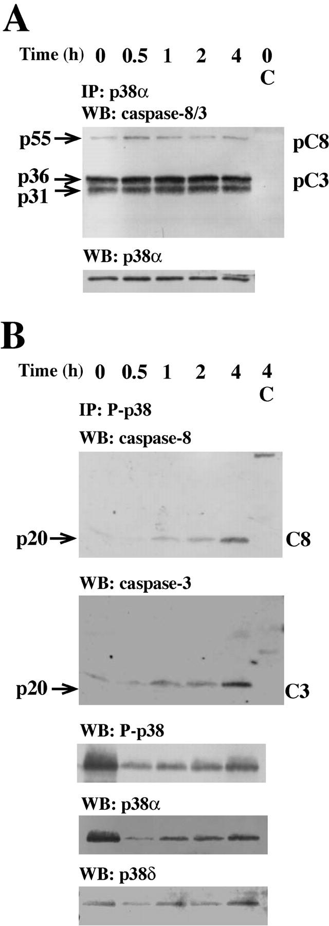
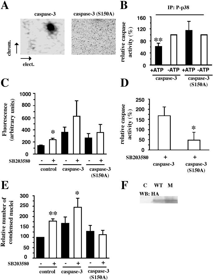
Neutrophil apoptosis occurs both in the bloodstream and in the tissue and is considered essential for the resolution of an inflammatory process. Here, we show that p38-mitogen-activated protein kinase (MAPK) associates to caspase-8 and caspase-3 during neutrophil apoptosis and that p38-MAPK activity, previously shown to be a survival signal in these primary cells, correlates with the levels of caspase-8 and caspase-3 phosphorylation. In in vitro experiments, immunoprecipitated active p38-MAPK phosphorylated and inhibited the activity of the active p20 subunits of caspase-8 and caspase-3. Phosphopeptide mapping revealed that these phosphorylations occurred on serine-364 and serine-150, respectively. Introduction of mutated (S150A), but not wild-type, TAT-tagged caspase-3 into primary neutrophils made the Fas-induced apoptotic response insensitive to p38-MAPK inhibition. Consequently, p38-MAPK can directly phosphorylate and inhibit the activities of caspase-8 and caspase-3 and thereby hinder neutrophil apoptosis, and, in so doing, regulate the inflammatory response.


Figures

Figure 1.
Figure 1.
Caspase-8 and caspase-3 are coimmunoprecipitated with p38-MAPK. Neutrophils were incubated with anti-Fas Ab for the indicated times and lysed. (A) Samples were immunoprecipitated with an anti–p38-MAPKα or an isotype-matched control (C) antibody and subsequently assessed by Western blotting. The blots were either analyzed with a mixture of two antibodies respectively directed against caspase-8 (both the proform, pC8, and the active form, C8) and caspase-3 (both the proform, pC3, and the active form, C3), sequentially stripped, and reprobed with antibodies against p38-MAPKα (p38α). (B) Samples were immunoprecipitated with an anti-phospho–p38-MAPK or an isotype-matched control (C) antibody and subsequently assessed by Western blotting. The blots were sequentially analyzed with the antibodies against caspase-8, caspase-3, phospho–p38-MAPK (P-p38), p38-MAPKα (p38α), and p38-MAPKδ (p38δ). The blots in A and B are representative of at least seven separate experiments.
Figure 2.
Figure 2.
p38-MAPK–dependent phosphorylations of procaspase-8 and procaspase-3 in intact cells. Neutrophils were incubated with anti-Fas Ab for the indicated times and lysed. (A) Samples were taken for Western blot analysis with an anti-phospho–p38-MAPK (P-p38) antibody. The blot shown is representative of at least eight separate experiments. (B) Alternatively, lysate samples were analyzed for IETDase (C8) and DEVDase (C3) activities (n = 6). To adjust for differences between blood batches, the caspase activities measured after 2 h were defined as 100%, and values at other time points were compared with that level. Caspase-3 immunoprecipitates were obtained from freshly isolated or 32P-labeled neutrophils after the indicated periods of exposure to anti-Fas Ab. (C) Unlabeled neutrophils were lysed, and the immunoprecipitates were immunoblotted with an anti–phospho-serine Ab, stripped, and reprobed with a mixture of anti–caspase-8 (detecting both the proform, pC8, and the active form) and anti–caspase-3 (detecting both the proform, pC3, and the active form) Abs and thereafter with an anti-phospho–p38-MAPK (P-p38) Ab. (D) The 32P-labeled neutrophils were lysed, and immunoprecipitates were analyzed by gel electrophoresis and blotted. The blots were developed with a PhosphorImager and subsequently analyzed with a mixture of the anti–caspase-8 and the anti–caspase-3 Abs, stripped, and reprobed with the anti-phospho–p38-MAPK Ab. The blots and the autoradiogram in C and D are representative of at least three separate experiments.
Figure 3.
Figure 3.
p38-MAPK–induced phosphorylations of caspase-8 and caspase-3 in vitro. (A and B) Active phosphorylated p38-MAPK immunoprecipitates from freshly isolated neutrophils were incubated with [γ-32P]ATP and recombinant caspase-8 or -3 in the absence or presence of the p38-MAPK inhibitor SB203580. As controls, the same reaction was performed in the absence of caspases but in the presence of either purified proteins from E. coli transformed with an empty vector (C) or BSA. All blots were first developed with a PhosphorImager, cut, and analyzed with (A) the anti–caspase-8 (pC8 and C8) Ab or (B) the anti–caspase-3 (pC3 and C3) Ab, and, lastly, stripped and reprobed with the anti-phospho–p38-MAPK (P-p38) Ab. The illustrated autoradiograms and blots are representative of at least seven separate experiments. (C) Active phosphorylated p38-MAPK (P-p38) was immunoprecipitated and incubated with recombinant caspase-8 and caspase-3 as substrates, in the presence or absence of ATP and under the same conditions as aforementioned. Thereafter, the activities of caspase-8 and caspase-3 were measured separately. The results are presented as percentage of the activities found in samples incubated in the same way but with an immunoprecipitate obtained using an isotype-matched control antibody. The data are expressed as mean ± SEM of seven separate experiments. The substrates, recombinant procaspase-8 (pC8) in D and recombinant procaspase-3 (pC3) in E, were incubated in the presence of ATP and either the immunoprecipitated active phosphorylated p38-MAPK (P-p38) or an immunoprecipitate obtained using an isotype-matched control antibody (Control). Thereafter, the in vitro amounts of the procaspases (pC8 and pC3) and caspases (C8 and C3), after incubations in the presence of (D) immunoprecipitated active Fas (FasR; n = 3) or (E) active caspase-8 (n = 5), were analyzed by Western blotting.
Figure 4.
Figure 4.
Identification of phosphorylation sites on caspase-8 and caspase-3. (A) Active phosphorylated p38-MAPK immunoprecipitates from freshly isolated neutrophils were incubated with [γ-32P]ATP and recombinant procaspase-8 or procaspase-3. The proteins were separated by SDS–gel electrophoresis, and the separated proteins were digested in situ with trypsin. The obtained phosphopeptides were separated on cellulose TLC glass plates (elect.), followed by ascending chromatography (chrom.). The indicated electrophoresis direction is from the anode to the cathode. The plates were analyzed in a PhosphorImager as well as exposed to an X-ray film. (B) The phosphopeptides from caspase-8 or caspase-3 were eluted from the TLC plates and subjected to two-dimensional phosphoamino acid analysis. The locations of the phosphoamino acids (top) were compared with that of phosphoamino acid markers (bottom) as follows: serine (S), threonine (T), and tyrosine (Y). The phosphopeptides obtained from A were subjected to amino acid sequencing (C), and the radioactivity released in each cycle was measured by spotting onto TLC plates and exposure on a Fuji image analyzer. The phosphorylated serine residues, 364 for caspase-8 (C) and 150 for caspase-3 (C), are indicated in the sequence of the putative fragment from caspase-8 and caspase-3, respectively. The illustrated phosphomapping is representative of three experiments.
Figure 5.
Figure 5.
Ser-364 and Ser-150 are conserved residues in caspases. (A) The homologous serine/threonine residue (first box) is found 11 amino acids upstream of the active site (second box) within the outlined caspases. These residues are found in the respective, large, p20 subunits of the caspases. (B) The known three dimensional structures of the p20 monomers of caspase-8 (34), caspase-3 (35), caspase-9 (36), and caspase-7 (37) are revealed with the three-dimensional structure viewer Cn3D. In these structures, the p38-MAPK putative phosphorylation site is depicted (yellow), below which is the active site of each caspase.
Figure 6.
Figure 6.
Mutation of Ser-150 abolishes the effect of the inhibition of p38-MAPK in vivo. (A) Active phosphorylated p38-MAPK immunoprecipitates from freshly isolated neutrophils were incubated with [γ-32P]ATP and recombinant wild-type TAT–caspase-3 or mutated TAT–caspase-3 (S150A). The phosphopeptide mapping was performed as in Fig. 4. The plates were exposed on a PhosphorImager as well as to film. The indicated electrophoresis direction is from the anode to the cathode. (B) Active phosphorylated p38-MAPK (P-p38) was immunoprecipitated and incubated with recombinant wild-type TAT–caspase-3 or mutated TAT-caspase-3 (S150A) as substrates in the presence (shaded bars) or absence (unshaded bars) of ATP. Thereafter, the activities of TAT–caspase-3 or TAT–caspase-3 (S150A) were measured separately. The results are presented as percentage of the activities found in samples depleted of ATP. The data are expressed as mean ± SD of five separate experiments. (C) Fas-induced caspase-3 activity in control cells or cells loaded with TAT–caspase-3 or TAT–caspase-3 (S150A) after 3 h after activation of the Fas-receptor. Unshaded bars represent cells treated with 20 μM SB203580 (n = 4–6). (D) The effect of SB203580 in cells loaded with TAT-fusion proteins (n = 4). (E) The Fas-treated cells were incubated for 4 h in the presence or absence of TAT-fusion protein and SB203580 and subsequently stained with acridine orange and ethidium bromide to assess their nuclear morphology. The effects are presented as percentage of untreated control cells (n = 4–6). (F) Neutrophils incubated in absence, as control (C) or presence of wild-type (WT) TAT-caspase-3 or (S150A) mutated (M) TAT–caspase-3. Samples were taken for Western blot analysis with an anti–HA-Ab (HA).

Similar articles

Cited by

References

    1. Cartwright, G.E., G.W. Athens, and M.M. Wintrobe. 1964. The kinetics of granulopoiesis in normal man. Blood. 24:780–803. - PubMed
    1. Savill, J.S., A.H. Wyllie, J.E. Henson, M.J. Walport, P.M. Henson, and C. Haslett. 1989. Macrophage phagocytosis of aging neutrophils in inflammation. Programmed cell death in the neutrophil leads to its recognition by macrophages. J. Clin. Invest. 83:865–875. - PMC - PubMed
    1. Grigg, J.M., J.S. Savill, C. Sarraf, C. Haslett, and M. Silverman. 1991. Neutrophil apoptosis and clearance from neonatal lungs. Lancet. 338:720–722. - PubMed
    1. Glogauer, M., J. Hartwig, and T. Stossel. 2000. Two pathways through Cdc42 couple the N-formyl receptor to actin nucleation in permeabilized human neutrophils. J. Cell Biol. 150:785–796. - PMC - PubMed
    1. Cryns, V., and J. Yuan. 1998. Proteases to die for. Genes Dev. 12:1551–1570. - PubMed

Publication types