. 2004 Feb 16;199(4):547-57.
doi: 10.1084/jem.20031685.

Deficiency of the cyclin kinase inhibitor p21(WAF-1/CIP-1) promotes apoptosis of activated/memory T cells and inhibits spontaneous systemic autoimmunity

Affiliations

Deficiency of the cyclin kinase inhibitor p21(WAF-1/CIP-1) promotes apoptosis of activated/memory T cells and inhibits spontaneous systemic autoimmunity

Brian R Lawson et al. J Exp Med. .

Abstract

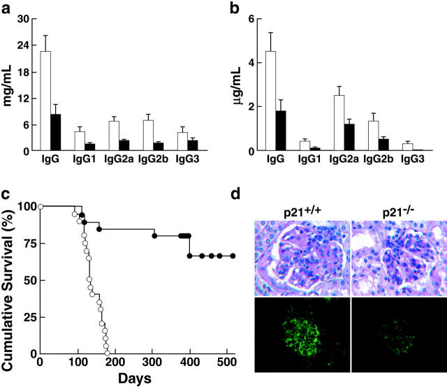
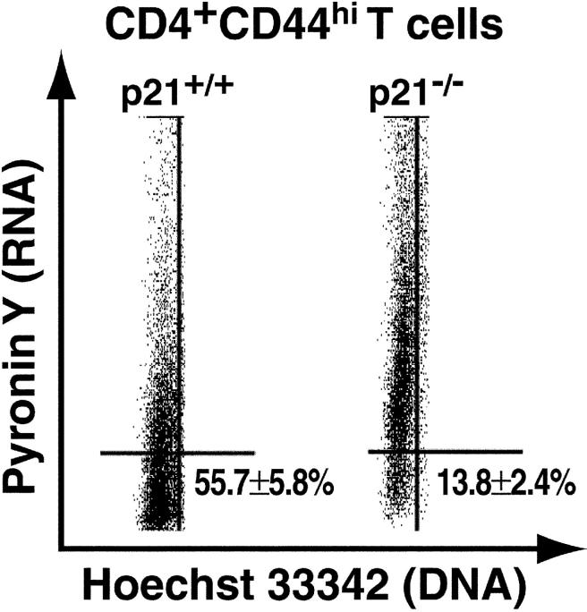
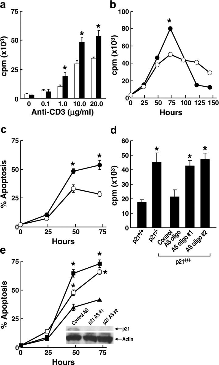
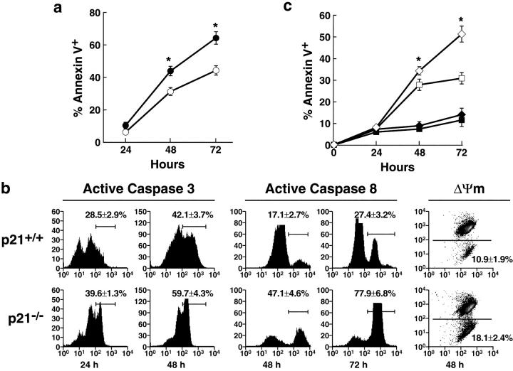
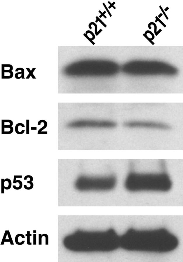
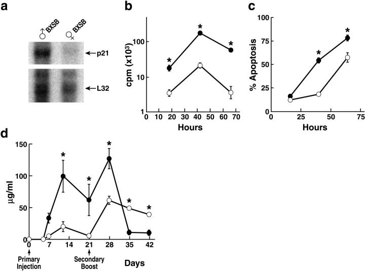
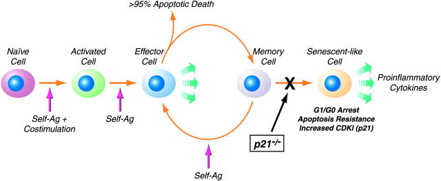
A characteristic feature of systemic lupus erythematosus is the accumulation of activated/memory T and B cells. These G0/G1-arrested cells express high levels of cyclin-dependent kinase inhibitors such as p21, are resistant to proliferation and apoptosis, and produce large amounts of proinflammatory cytokines. Herein, we show that ablation of p21 in lupus-prone mice allows these cells to reenter the cell cycle and undergo apoptosis, leading to autoimmune disease reduction. Absence of p21 resulted in enhanced Fas/FasL-mediated activation-induced T cell death, increased activation of procaspases 8 and 3, and loss of mitochondrial transmembrane potential. Increased apoptosis was also associated with p53 up-regulation and a modest shift in the ratio of Bax/Bcl-2 toward the proapoptotic Bax. Proliferation and apoptosis of B cells were also increased in p21-/- lupus mice. Thus, modulation of the cell cycle pathway may be a novel approach to reduce apoptosis-resistant pathogenic lymphocytes and to ameliorate systemic autoimmunity.


Figures

Figure 1.
Figure 1.
Decreased autoimmune disease in male p21−/− BXSB mice. (a and b) Reduced serum polyclonal (left) and antichromatin (right) IgG subclasses in 4-mo-old male BXSB p21−/− mice (n = 8 mice/group; mean ± SEM). (shaded bars) BXSB p21−/− mice. (unshaded bars) p21+/+ BXSB mice. For all polyclonal and antichromatin total and subclass IgG levels, P < 0.05 for p21−/− versus p21+/+. (c) Increased cumulative survival rates of p21−/− BXSB mice (P < 0.0001). Male BXSB p21+/+ (n = 16) and p21−/− (n = 15) littermates were followed for up to 500 d. (•) BXSB p21−/− mice. (○) BXSB p21+/+ mice. (d) Glomerular pathology (top) and IgG deposits (bottom) of representative 4-mo-old p21+/+ and p21−/− mice. Increased segmental mesangial proliferation and accumulation of periodic-acid Schiff–positive mesangial matrix material were seen in p21+/+ mice, whereas p21−/− mice exhibited significantly less glomerular damage, as well as decreased segmental granular mesangial and capillary wall deposits of IgG (top, 630×; bottom, 400×).
Figure 2.
Figure 2.
Representative distribution of G0- versus G1-phase in CD4+CD44hi T cells. Splenocytes from 3-mo-old mice (n = 4 animals/group) were stained with antibodies to CD4 and CD44, followed by sequential incubation with Hoechst 33342 (DNA-binding dye) and Pyronin Y (RNA-binding dye) and analyzed by flow cytometry. Pyronin Y staining of G0/G1-phase CD4+CD44hi T cells from p21−/− and p21+/+ BXSB mice is shown (P < 0.05).
Figure 3.
Figure 3.
Enhanced proliferation and apoptosis of p21−/− T cells from male BXSB mice. (a) Increased proliferation of p21−/− T cells after in vitro cross-linking of antigen receptors. LN cells (n = 3 mice/group) were stimulated with increasing concentrations of plate-bound anti-CD3 (0.1–20 μg/ml) plus 5 μg/ml of soluble anti-CD28 antibodies and assessed for [3H]thymidine incorporation at 72 h (mean ± SEM cpm). (shaded bars) BXSB p21−/− mice. (unshaded bars) p21+/+ BXSB mice. (b) Kinetics of in vitro proliferation. LN cells (n = 3 mice/group) were stimulated with 10 μg/ml of plate-bound anti-CD3 plus 5 μg/ml of soluble anti-CD28 antibodies and assessed for [3H]thymidine incorporation at the indicated time points. Results are representative of two independent experiments (n = 3 mice/group). (•) BXSB p21−/− mice. (○) BXSB p21+/+ mice. (c) Increased AICD of p21−/− T cells. T cell were stimulated with 0.5 μg/ml of soluble anti-CD3 for 48 h followed by TCR religation with 10 μg/ml of plate-bound anti-CD3 (n = 3 mice/group). Percent of apoptotic (Annexin V+PI) CD4+ T cells was determined by FACS®. Similar results were obtained with CD8+ T cells. (•) BXSB p21−/− mice. (○) BXSB p21+/+ mice. (d) Increased proliferation of wild-type BXSB T cells treated with p21 AS oligonucleotides. LN cells (n = 4 mice/group) were stimulated with 10 μg/ml of plate-bound anti-CD3 plus 5 μg/ml of soluble anti-CD28 antibodies in the presence of either p21 AS or control oligonucleotides (all AS oligonucleotides at 400 nM) and assessed for [3H]thymidine incorporation (mean ± SEM cpm). (e) Increased AICD of wild-type BXSB T cells treated with p21 AS oligonucleotides. Wild-type BXSB T cells were stimulated with 0.5 μg/ml of soluble anti-CD3 followed by TCR religation with 10 μg/ml of plate-bound anti-CD3 in the constant presence of p21 AS or control oligonucleotides (400 nM; n = 3 mice/group). (▪) AS oligo no. 1. (□) AS oligo no. 2. (▴) Control oligonucleotide. (e, inset) Western blot analysis of wild-type BXSB T cells treated with either p21 AS or control oligonucleotides. T cells were stimulated with 10 μg/ml anti-CD3 and 5 μg/ml CD28 antibodies in the presence of AS or control oligonucleotides (400 nm). Whole cell lysates were analyzed by Western blot using anti-p21 and antiactin antibodies. *, P < 0.05 by Student's t test.
Figure 4.
Figure 4.
Enhanced Fas/FasL-mediated apoptosis of p21−/− T cells from male BXSB mice. (a) Increased anti-Fas induced apoptosis of p21−/− T cells. LN cells (n = 3 mice/group) were first stimulated with 10 μg/ml of soluble anti-CD3 and 5 μg/ml CD28 for 48 h and then with 5 μg/ml anti-Fas antibody and analyzed for percentage of apoptotic (Annexin V+PI) CD4+ T cells. (•) BXSB p21−/− mice. (○) BXSB p21+/+ mice. *, P < 0.05. (b) Increased caspase 8 and 3 activation and reduced mitochrondrial membrane potential (ΔΨm) in p21−/− T cells after induction of AICD. T cells (n = 3 mice/group) were stimulated with 0.1 μg/ml of soluble anti-CD3, religated with 10 μg/ml of plate-bound anti-CD3, and analyzed by FACS® for activation of caspases 8 and 3 and for change in mitochondrial transmembrane potential. The percentage of cells expressing activated caspases 8 or 3 and reduced ΔΨm is indicated (mean ± SEM). P < 0.05 between p21+/+ and p21−/− for all time points shown. (c) Fas blockade inhibits AICD of p21+/+ and p21−/− T cells. T cells (n = 3 mice/group) were stimulated with 0.1 μg/ml of soluble anti-CD3 for 48 h, religated with 10 μg/ml of plate-bound anti-CD3 in the presence or absence of 10 μg/ml of blocking anti-FasL antibody, and analyzed for Annexin V positivity. Similar inhibition was also observed after treatment with Fas-blocking soluble Fas/Fc. (♦) BXSB p21−/− mice plus anti-FasL. (▪) BXSB p21+/+ mice plus anti-FasL. (⋄) BXSB p21−/− mice. (□) BXSB p21+/+ mice. *, P < 0.05 for untreated (⋄ or □) versus anti-FasL–treated groups (♦ or ▪) at 48 and 72 h, and for untreated p21−/− (⋄) versus p21+/+ (□) mice at 72 h.
Figure 5.
Figure 5.
Participation of the intrinsic pathway of apoptosis in p21−/− T cells from male BXSB mice. Increased p53 and reduced Bcl-2 expression in activated p21−/− T cells. T cells (n = 5 mice/group) were activated with 10 μg/ml anti-CD3 plus 5 μg/ml anti-CD28, and lysates were analyzed by Western blot.
Figure 6.
Figure 6.
Enhanced proliferation and apoptosis, and reduced late secondary immunoglobulin responses of p21−/− B cells from male BXSB mice. (a) Increased p21 expression in B cells from older male BXSB mice. Sorted B cells (CD19+) from 3-mo-old wild-type male or female BXSB mice (n = 5 mice/group) were analyzed by RNase protection assay for expression of p21 and L32 (control). (b) Increased proliferation of p21−/− B cells after IgM cross-linking. Splenocytes (n = 3 mice/group) were stimulated with 10 μg/ml of soluble goat F(ab′)2 anti–mouse IgM in the presence of IL-4 and assessed for [3H]thymidine incorporation (mean ± SEM cpm). (•) BXSB p21−/− mice. (○) BXSB p21+/+ mice. (c) Enhanced AICD of p21−/− B cells. Annexin V positivity of B cells was assessed after anti-IgM stimulation with 10 μg/ml of soluble goat F(ab′)2 anti–mouse IgM (n = 3 mice/group). (•) BXSB p21−/− mice. (○) BXSB p21+/+ mice. (d) Anti-TNP antibody levels after primary and secondary immunizations. 2-mo-old male BXSB p21−/− and p21+/+ mice (n = 4 mice/group) were injected s.c. with 100 μg TNP-KLH emulsified in CFA. Secondary responses were assessed by boosting mice s.c. with 100 μg TNP-KLH in saline on day 21. Mice were bled at the indicated times, and serum was analyzed by ELISA. (•) BXSB p21−/− mice. (○) BXSB p21+/+ mice. *, P < 0.05 by Student's t test.
Figure 7.
Figure 7.
T cell senescence and p21 in systemic autoimmunity. In lupus-prone mice, the accumulated activated/memory CD4+ T cells are in a state resembling replicative senescence. We hypothesize that repeated stimulation of CD4+ T cells (depicted by the hypothetical movement of cells from the memory to effector compartments) by self-antigens (Ag) leads to resistance to proliferation and apoptosis (senescent-like cell), due in large part to increased levels of CDKIs. Senescent cells are metabolically active and can produce proinflammatory cytokines. As shown in this work, the gradual accumulation of these activated/memory phenotype CD4+ T cells and subsequent development of autoimmunity is dependent on p21. Furthermore, our data indicate that the fraction of activated/memory CD4+CD44hi T cells that escape AICD in wild-type autoimmune mice (because of increased CDKIs) do not accumulate in G0/G1-phase in p21-deficient mice; instead, they proliferate and become susceptible to Fas-mediated apoptosis. Thus, the lack of p21 appears to restore homeostasis of autoreactive CD4+CD44hi T cells by preventing their transition to a senescent-like state.

Similar articles

Cited by

References

    1. Sprent, J., and D.F. Tough. 1994. Lymphocyte life-span and memory. Science. 265:1395–1400. - PubMed
    1. Ahmed, R., and D. Gray. 1996. Immunological memory and protective immunity: understanding their relation. Science. 272:54–60. - PubMed
    1. Theofilopoulos, A.N., and D.H. Kono. 1999. Murine lupus models: gene-specific and genome-wide studies. Systemic Lupus Erythematosus. R.G. Lahita, editor. Academic Press, San Diego. 145–181.
    1. Chu, E.B., D.N. Ernst, M.V. Hobbs, and W.O. Weigle. 1994. Maturational changes in CD4+ cell subsets and lymphokine production in BXSB mice. J. Immunol. 152:4129–4138. - PubMed
    1. Sabzevari, H., S. Propp, D.H. Kono, and A.N. Theofilopoulos. 1997. G1 arrest and high expression of cyclin kinase and apoptosis inhibitors in accumulated activated/memory phenotype CD4+ cells of older lupus mice. Eur. J. Immunol. 27:1901–1910. - PubMed

Publication types