Review
. 2004 Mar;10(3):335-43.
doi: 10.1261/rna.5205404.

A guide to ions and RNA structure

Affiliations
Review

A guide to ions and RNA structure

David E Draper. RNA. 2004 Mar.

Abstract

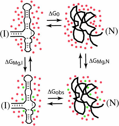
RNA folding into stable tertiary structures is remarkably sensitive to the concentrations and types of cations present; an understanding of the physical basis of ion-RNA interactions is therefore a prerequisite for a quantitative accounting of RNA stability. This article summarizes the energetic factors that must be considered when ions interact with two different RNA environments. "Diffuse ions" accumulate near the RNA because of the RNA electrostatic field and remain largely hydrated. A "chelated" ion directly contacts a specific location on the RNA surface and is held in place by electrostatic forces. Energetic costs of ion chelation include displacement of some of the waters of hydration by the RNA surface and repulsion of diffuse ions. Methods are discussed for computing both the free energy of the set of diffuse ions associated with an RNA and the binding free energies of individual chelated ions. Such calculations quantitatively account for the effects of Mg(2+) on RNA stability where experimental data are available. An important conclusion is that diffuse ions are a major factor in the stabilization of RNA tertiary structures.


Figures

FIGURE 1.
FIGURE 1.
Potential ion environments in an RNA. Represented are a Mg2+ ion (green dot), nonbridging phosphate oxygens (red circles at the hatched RNA surface), and water molecules (red and gray). At the left, Mg2+ is chelated and partially dehydrated by RNA phosphates; at the right, the Mg2+ remains hydrated but still interacting with the RNA electrostatic field. A possible intermediate situation, in which Mg2+ retains one layer of hydrating water molecules that in turn interact with the RNA surface, is shown in the middle.
FIGURE 2.
FIGURE 2.
Distribution of ions in equilibrium dialysis experiments with RNA. (A) When an RNA is dialyzed against a KCl solution, K+ (blue) accumulates inside the dialysis bag and Cl (yellow) is excluded from the bag, relative to the concentrations of the ions outside the bag. Quantitation of the retained and excluded ions is shown in the series of histograms BE (y-axis: K+, Cl, and phosphate ion concentrations inside or outside the dialysis bag). (B) “Initial” shows the concentrations of ions after the RNA is placed in the dialysis bag but before dialysis begins. (C) “Ideal” shows the equilibrium concentrations that would be reached if the phosphate charges were infinitely far apart. (D,E) Show the ion concentrations for poly(U) (0.84 K+ retained per phosphate) or poly(U)•poly(A) (0.94 K+ retained per phosphate). The quantitations are based on polyelectrolyte theory and are valid in the limit of low salt concentration, although the cited numbers are relatively independent of the “outside” KCl concentration over a wide range. This diagram and the quantitations of ion retentions are adapted from Record and Richey (1988).
FIGURE 3.
FIGURE 3.
The ion atmosphere around an RNA helix. Red shading, the electrostatic potential; the contour lines map the concentration of cations. Contour lines range from 1.6 M (brown) to 0.2 M (magenta) with intermediate concentrations of 0.8 M and 0.4 M. The bulk monovalent salt concentration is 0.1 M. The highest concentration of cations is found in the major groove, where the electrostatic potential is also highest. Based on Figure 6 of García-García and Draper (2003).
FIGURE 4.
FIGURE 4.
Energetics of placing a Mg2+ ion in an RNA chelation site. (A,B) The major unfavorable energetic factors. The ion must be partially dehydrated to make direct contact with the RNA surface (ΔGhyd, panel A). The chelated ion displaces ~two diffuse ions from the ion atmosphere, removing their favorable interactions with the RNA, and has repulsive interactions (gray arrows) with the remaining diffuse ions (ΔGMg-diffuse, panel B). Mg2+ interactions with the RNA are favorable (double-headed arrows) and almost entirely electrostatic (ΔGMg-RNA, panel C). Only in unusual chelation sites is ΔGMg-RNA large enough to outweigh the other two unfavorable factors.
FIGURE 5.
FIGURE 5.
Thermodynamic cycle used to relate the measurement of Mg2+-induced stabilization of an RNA tertiary structure to calculations of Mg2+ interactions with RNA. The two left-hand diagrams represent an rRNA fragment with only secondary structure present (the partially folded I state); the right-hand structures are the same RNA in its native tertiary structure (N state). Red dots represent K+ accumulated by the RNA in excess of the K+ concentration in the bulk solution; green dots are Mg2+ added in the reactions depicted by the vertical arrows. (The depletion of anions in the vicinity of the RNA is not illustrated.) The overall free energy in going from I state RNA in the absence of Mg2+ (upper left) to the native RNA with Mg2+ (lower right) is the same whichever pathway is taken; thus ΔG0 + ΔGMg,N = ΔGMg,I + ΔGobs. Mg2+-induced stabilization of the RNA tertiary structure is the difference between the stabilities in the presence and absence of Mg2+, ΔGobs − ΔG0. By the foregoing constraint, this must be equal to ΔGMg,N − ΔGMg,I, a quantity that can be calculated by the approaches described in the text.

References

    1. Amzel, L.M. 1997. Loss of translational entropy in binding, folding, and catalysis. Proteins 28: 144–149. - PubMed
    1. Anderson, C.F. and Record Jr., M.T. 1995. Salt-nucleic acid interactions. Annu. Rev. Phys. Chem. 46: 657–700. - PubMed
    1. Basu, S. and Strobel, S.A. 1999. Thiophilic metal ion rescue of phosphorothioate interference within the Tetrahymena ribozyme P4–P6 domain. RNA 5: 1399–1407. - PMC - PubMed
    1. Bokinsky, G., Rueda, D., Misra, V.K., Rhodes, M.M., Gordus, A., Babcock, H.P., Walter, N.G., and Zhuang, X. 2003. Single-molecule transition-state analysis of RNA folding. Proc. Natl. Acad. Sci. 100: 9302–9307. - PMC - PubMed
    1. Braunlin, W.H. 1995. NMR Studies of cation-binding environments on nucleic acids. Adv. Biophys. Chem. 5: 89–139.

Publication types

LinkOut - more resources