. 2004 Mar;10(3):378-86.
doi: 10.1261/rna.5169404.

Nascent-peptide-mediated ribosome stalling at a stop codon induces mRNA cleavage resulting in nonstop mRNA that is recognized by tmRNA

Affiliations

Nascent-peptide-mediated ribosome stalling at a stop codon induces mRNA cleavage resulting in nonstop mRNA that is recognized by tmRNA

Takafumi Sunohara et al. RNA. 2004 Mar.

Abstract

Recent studies have established that tmRNA-mediated protein tagging occurs at stop codons depending on the C-terminal amino acid sequence of the nascent polypeptide immediately adjacent to those codons. We investigate here how the trans-translation at a stop codon occurs by using model crp genes encoding variants of cAMP receptor protein (CRP). We demonstrate that a truncated crp mRNA is efficiently produced along with a normal transcript from the model gene where tmRNA-mediated protein tagging occurs. The truncated crp mRNA was not detected in the presence of tmRNA, indicating that its degradation was facilitated by tmRNA. The major 3'-ends of the truncated crp mRNA in cells unable to express tmRNA were mapped at and near the stop codon. When RNA derived from the model crp-crr fusion gene was analyzed, crr mRNA was detected as a downstream cleavage product along with the upstream crp mRNA. These results are compatible with the hypothesis that ribosome stalling caused by the tagging-provoking sequences leads to endonucleolytic cleavage of mRNA around the stop codon, resulting in nonstop mRNA. In addition, the data are consistent with the view that mRNA cleavage is the cause of trans-translation at stop codons. Neither the bacterial toxin RelE nor the known major endoribonucleases are required for this cleavage, indicating that either other endoribonuclease(s) or the ribosome itself would be responsible for the mRNA cleavage in response to ribosome stalling caused by the particular nascent peptides.


Figures

FIGURE 1.
FIGURE 1.
Schematic drawing of the (A) crp and (B) crpcrr fusion genes used in this study. The open and shaded rectangles represent the coding region for CRP and IIAGlc, respectively. The black box represents the altered 3′-portion of the CRP coding region. The nucleotide sequence and amino acid sequence (in one-letter symbols) of the variable region are shown below the diagram.
FIGURE 2.
FIGURE 2.
Tagging of a series of CRP-XP proteins. Lysates equivalent to OD600 = 0.005 unit prepared from TA481 (Δcrp ssrADD) cells harboring pHA7 derivatives carrying altered crp genes that encode CRP-XP proteins were analyzed by Western blotting using anti-CRP antibodies. The amino acid residues at the −2 position are indicated by a one-letter symbol. The −2 and −1 codons were (A) GCG CCG; (C) TGC CCG; (D) GAT CCA; (E) GAA CCG; (F) TTC CCC; (G) GGC CCT; (H) CAT CCG; (I) ATT CCG; (K) AAA CCG; (L) CTG CCG; (M) ATG CCG; (N) AAC CCG; (P) CCA CCT; (Q) CAG CCG; (R) CGC CCG; (S) AGC CCG; (T) ACC CCG; (V) GTG CCT; (W) TGG CCG; and (Y) TAT CCG.
FIGURE 3.
FIGURE 3.
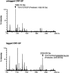
Mass spectrometry analysis of untagged and tagged CRP-GP. Purified proteins were separated on a 12% SDS-polyacrylamide gel electrophoresis followed by Coomassie Brilliant Blue staining. The bands corresponding to untagged and tagged CRP-GP were cut out from the gel. The gel was treated with lysyl endopeptidase and subjected to mass spectrometry analysis. The signals that are expected to correspond to the C-terminal fragments are shown by arrowheads along with the observed mass. The expected peptide sequences and molecular weights of the C-terminal fragments of untagged (upper) and tagged CRP-GP (lower) generated by lysyl endopeptidase digestion are shown below the observed mass.
FIGURE 4.
FIGURE 4.
Positional effect of the tagging-provoking sequences. Lysates equivalent to OD600 = 0.005 unit prepared from TA341 (Δcrp ssrA+), TA501 (Δcrp ΔssrA), or TA481 (Δcrp ssrADD) cells harboring indicated plasmids were analyzed by Western blotting using anti-CRP antibodies.
FIGURE 5.
FIGURE 5.
Northern blot analysis of crp mRNAs derived from the crp genes encoding CRP variants. Total RNA was prepared from TA341 (Δcrp ssrA+) and TA501 (Δcrp ΔssrA) cells harboring indicated plasmids. Either 0.15 μg (lanes 1,2,3,5,7,8) or 0.5 μg (lanes 4,6) of RNA was resolved by electrophoresis on a 2.0% agarose-formaldehyde gel. Northern blot analysis was performed using the DIG-labeled crp probe. RNA bands corresponding to the full-length and truncated crp mRNAs are indicated by arrowheads.
FIGURE 6.
FIGURE 6.
Determination of 3′-ends of the crp mRNAs. Total RNA (50 μg) prepared from TA501 (Δcrp ΔssrA) harboring pJK021 was hybridized with the DNA probe C 32P-labeled at its 3′-end of the template strand, and the hybrids were treated with the indicated amounts of S1 nuclease. The products were dissolved in 20 μL of loading buffer (8 M urea, 0.025% bromophenol blue, 0.025% xylene cyanol, 90 mM Tris-borate at pH 8.3, and 1 mM EDTA), and 2 μL of each sample was analyzed on an 8% polyacrylamide–8 M urea gel along with products of an A + G and C + T chemical sequencing reaction of the fragment. Cluster I represents the 3′-ends of the normal crp mRNA, whereas cluster II corresponds to the truncated crp mRNA. The nucleotide sequence around the stop codon of the altered crp gene is shown on the right. The arrowheads indicate the major 3′-ends identified by the S1 analysis. The TAA stop codon is underlined. The GC-rich inverted repeat sequence of the terminator is indicated by vertical arrows.
FIGURE 7.
FIGURE 7.
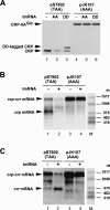
Analyses of protein and RNA derived from the crpcrr fusion genes. (A) Western blot analysis of CRP proteins. Lysates equivalent to OD600 = 0.005 unit prepared from TA341 (Δcrp ssrA+), TA501 (Δcrp ΔssrA), or TA481 (Δcrp ssrADD) cells harboring pST602 (lanes 13) and pJK107 (lanes 46) were analyzed by Western blotting using anti-CRP antibodies. (B) Northern blot analysis of crp mRNAs derived from the crpcrr fusion genes. Total RNA (1 μg) prepared from TA341 (Δcrp ssrA+) and TA501 (Δcrp ΔssrA) cells harboring pST602 (lanes 1,2) and pJK107 (lanes 3,4) were resolved by electrophoresis on a 1.5% agarose-formaldehyde gel. Northern blot analysis was performed using the DIG-labeled crp probe. RNA bands corresponding to the full-length crpcrr and truncated crp mRNAs are indicated by arrowheads. (Lane M) RNA size markers. (C) Northern blot analysis of crr mRNAs derived from the crpcrr fusion genes. Total RNA (1 μg) prepared from TA341 (Δcrp ssrA+) and TA501 (Δcrp ΔssrA) cells harboring pST602 (lanes 1,2) and pJK107 (lanes 3,4) were resolved by electrophoresis on a 1.5% agarose-formaldehyde gel. Northern blot analysis was performed using the DIG-labeled crr probe. RNA bands corresponding to the full-length crpcrr and truncated crr mRNAs are shown by arrowheads. (Lane M) RNA size markers.
FIGURE 8.
FIGURE 8.
Effect of relEB disruption on mRNA cleavage. (A) Northern blot analysis of crp mRNAs derived from the crpcrr fusion genes. Total RNA (1 μg) prepared from ST100 (ΔrelEB) and ST101 (ΔssrA ΔrelEB) cells harboring pST602 (lanes 1,2) and pJK107 (lanes 3,4) were resolved by electrophoresis on a 1.5% agarose-formaldehyde gel. Northern blot analysis was performed using the DIG-labeled crp probe. RNA bands corresponding to the full-length crpcrr and truncated crp mRNAs are indicated by arrowheads. (Lane M) RNA size markers. (B) Northern blot analysis of crr mRNAs derived from the crpcrr fusion genes. Total RNA (1 μg) prepared from ST100 (ΔrelEB) and ST101 (ΔssrA ΔrelEB) cells harboring pST602 (lanes 1,2) and pJK107 (lanes 3,4) were resolved by electrophoresis on a 1.5% agarose-formaldehyde gel. Northern blot analysis was performed using the DIG-labeled crr probe. RNA bands corresponding to the full-length crpcrr and truncated crr mRNAs are indicated by arrowheads. (Lane M) RNA size markers.
FIGURE 9.
FIGURE 9.
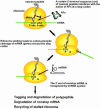
Model for mRNA cleavage at a stop codon induced by ribosome stalling.

References

    1. Abe, H., Abo, T., and Aiba, H. 1999. Regulation of intrinsic terminator by translation in Escherichia coli: Transcription termination at a distance downstream. Genes Cells 4: 87–97. - PubMed
    1. Abo, T., Inada, T., Ogawa, K., and Aiba, H. 2000. SsrA-mediated tagging and proteolysis of LacI and its role in the regulation of lac operon. EMBO J. 19: 3762–3769. - PMC - PubMed
    1. Abo, T., Ueda, K., Sunohara, T., Ogawa, K., and Aiba, H. 2002. SsrA-mediated protein tagging in the presence of miscoding drugs and its physiological role in Escherichia coli. Genes Cells 7: 629–638. - PubMed
    1. Aiba, H., Adhya, S., and de Crombrugghe, B. 1981. Evidence for two functional gal promoters in intact Escherichia coli cells. J. Biol. Chem. 256: 11905–11910. - PubMed
    1. Aiba, H., Fujimoto, S., and Ozaki, N. 1982. Molecular cloning and nucleotide sequencing of the gene for E. coli cAMP receptor protein. Nucleic Acids Res. 10: 1345–1361. - PMC - PubMed

Publication types

MeSH terms