. 2004 Mar;10(3):482-92.
doi: 10.1261/rna.5163104.

Interaction of the Bacillus subtilis RNase P with the 30S ribosomal subunit

Affiliations

Interaction of the Bacillus subtilis RNase P with the 30S ribosomal subunit

Alessandra Barrera et al. RNA. 2004 Mar.

Abstract

Ribonuclease P (RNase P) is a ribozyme required for the 5' maturation of all tRNA. RNase P and the ribosome are the only known ribozymes conserved in all organisms. We set out to determine whether this ribonucleoprotein enzyme interacts with other cellular components, which may imply other functions for this conserved ribozyme. Incubation of the Bacillus subtilis RNase P holoenzyme with fractionated B. subtilis cellular extracts and purified ribosomal subunits results in the formation of a gel-shifted complex with the 30S ribosomal subunit at a binding affinity of approximately 40 nM in 0.1 M NH(4)Cl and 10 mM MgCl(2). The complex does not form with the RNase P RNA alone and is disrupted by a mRNA mimic polyuridine, but is stable in the presence of high concentrations of mature tRNA. Endogenous RNase P can also be detected in the 30S ribosomal fraction. Cleavage of a pre-tRNA substrate by the RNase P holoenzyme remains the same in the presence of the 30S ribosome, but the cleavage of an artificial non-tRNA substrate is inhibited eightfold. Hydroxyl radical protection and chemical modification identify several protected residues located in a highly conserved region in the RNase P RNA. A single mutation within this region significantly reduces binding, providing strong support on the specificity of the RNase P-30S ribosome complex. Our results also suggest that the dimeric form of the RNase P is primarily involved in 30S ribosome binding. We discuss several models on a potential function of the RNase P-30S ribosome complex.


Figures
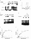
FIGURE 1.
FIGURE 1.
(A) Native gel analysis for RNase P binding with various fractions of B. subtilis extract. The holoenzyme dimer and monomer are well separated, but the dissociation of the dimer during gel electrophoresis generates a smear between the dimer and the monomer. A small fraction of the P RNA (<5%) also dimerizes in the absence of the P protein; such a dimerization of P RNA alone is due to the formation of an intermolecular P1 helix (X. Fang and T. Pan, unpubl. results). A severely retarded band is present with the S30 fraction, and incubation with the S100 fraction only generates a minor well shift. (B) Sucrose gradient of the 70S ribosomal fraction (dissolved S100 pellet) showing the separation of the pooled 30S and 50S ribosomal fractions. Two thick lines indicate the pooled fractions. (C) Gel shift of the RNase P holoenzyme by the purified ribosomal fractions. (D) Competition assay in the presence of 0.2 μg/μL Poly-U RNA, 5 μM yeast tRNAPhe, and 5 μM pre-tRNAPhe substrate (“tRNAPhe”). Because 30S binding does not affect the catalytic activity of the RNase P holoenzyme on this pre-tRNA substrate, it was certain that all pre-tRNA substrates had been cleaved before the sample was loaded onto the native gel. (E) Percent gel-shifted complex as a function of 30S ribosome concentration using the RNase P holoenzyme (filled circles) and a P RNA fragment containing residues 1–239 plus stoichiometric amount of P protein (open circles). (F) Percent gel-shifted complex as a function of 30S ribosome concentration in the presence of 0.1 mg/mL E. coli tRNA (type XXI from Sigma-Aldrich).
FIGURE 2.
FIGURE 2.
Detection of endogenous RNase P that copurifies with the ribosomal fractions by primer extension. Two product bands are detected in the 30S fraction. The shorter product probably is derived from removal of one or two 5′-terminal nucleotides in the P RNA during 30S purification. The single-stranded nt 1–4 in the P RNA are dispensable for all known functions of RNase P. The minor products in the 50S fraction are either derived from the 30S impurity in the pooled 50S fraction (see Fig. 1B ▶) or are unidentified reverse transcription products.
FIGURE 3.
FIGURE 3.
(A) Hydroxyl radical protection of the RNase P–30S ribosome complex. A thick line on the right indicates the protected region. (B) DMS and kethoxal modification of the RNase P-30S ribosome complex. (Left) Reverse transcriptase sequencing using the primer complementary to residues 367–351 of the P RNA; (middle) primer extension with the 367–351 complementary primer; (right) primer extension with the 402–381 complementary primer. A thick line on the right indicates the protected residues. (C) Summary of the protection and chemical modification results. Residues protected against hydroxyl radical attack are enclosed by an oval. Residues protected against DMS modification are shaded. The four regions involved in holoenzyme dimerization are shown as I–IV. Regions I, II, III, and IV include residues 107–116, 157–161, 198–223, and 264–273, respectively. Residues not examined for hydroxyl radical protection due to gel resolution are shown in lowercase.
FIGURE 4.
FIGURE 4.
(A) The extent of the protection of the four regions involved in the formation of the RNase P holoenzyme dimer in the RNase P–30S ribosome complex. For each position, the amount of radioactivity in the absence of 30S subunit divided by the amount of radioactivity in the presence of 30S subunit is shown as closed squares. The amount of radioactivity of the RNase P monomer divided by the amount of radioactivity of the RNase P dimer is shown as open circles (data taken from Barrera et al. 2002). (B) The inverse of the extent of protection versus 30S ribosome concentration can be fit to obtain an estimated binding affinity.
FIGURE 5.
FIGURE 5.
Native gel analysis of mutant holoenzymes. The RNase P concentration was 0.2 μM in all cases.

References

    1. Alifano, P., Rivellini, F., Piscitelli, C., Arraiano, C.M., Bruni, C.B., and Carlomagno, M.S. 1994. Ribonuclease E provides substrates for ribonuclease P-dependent processing of a polycistronic mRNA. Genes & Dev. 8: 3021–3031. - PubMed
    1. Altman, S. and Kirsebom, L. 1999. Ribonuclease P. In The RNA world, 2nd ed. (eds. R.F. Gesteland et al.), pp. 351–380. Cold Spring Harbor Laboratory Press, Cold Spring Harbor, NY.
    1. Barrera, A., Fang, X., Jacob, J., Casey, E., Thiyagarajan, P., Pan, T. 2002. Dimeric and monomeric Bacillus subtilis RNase P holoenzyme in the absence and presence of pre-tRNA substrates. Biochemistry 41: 12986–12994. - PubMed
    1. Brown, J.W. 1999. The Ribonuclease P Database. Nucleic Acids Res. 27: 314. - PMC - PubMed
    1. Chen, J.L., Nolan, J.M., Harris, M.E., and Pace, N.R. 1998. Comparative photocross-linking analysis of the tertiary structures of Escherichia coli and Bacillus subtilis RNase P RNAs. EMBO J. 17: 1515–1525. - PMC - PubMed

Publication types

MeSH terms

LinkOut - more resources