Monitoring temozolomide treatment of low-grade glioma with proton magnetic resonance spectroscopy
- PMID: 14970853
- PMCID: PMC2410174
- DOI: 10.1038/sj.bjc.6601593
Monitoring temozolomide treatment of low-grade glioma with proton magnetic resonance spectroscopy
Abstract
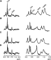
Assessment of low-grade glioma treatment response remains as much of a challenge as the treatment itself. Proton magnetic resonance spectroscopy ((1)H-MRS) and imaging were incorporated into a study of patients receiving temozolomide therapy for low-grade glioma in order to evaluate and monitor tumour metabolite and volume changes during treatment. Patients (n=12) received oral temozolomide (200 mg m(-2) day(-1)) over 5 days on a 28-day cycle for 12 cycles. Response assessment included baseline and three-monthly magnetic resonance imaging studies (pretreatment, 3, 6, 9 and 12 months) assessing the tumour size. Short (TE (echo time)=20 ms) and long (TE=135 ms) echo time single voxel spectroscopy was performed in parallel to determine metabolite profiles. The mean tumour volume change at the end of treatment was -33% (s.d.=20). The dominant metabolite in long echo time spectra was choline. At 12 months, a significant reduction in the mean choline signal was observed compared with the pretreatment (P=0.035) and 3-month scan (P=0.021). The reduction in the tumour choline/water signal paralleled tumour volume change and may reflect the therapeutic effect of temozolomide.
Figures


References
-
- Barba I, Cabanas ME, Arus C (1999) The relationship between nuclear magnetic resonance-visible lipids, lipid droplets, and cell proliferation in cultured C6 cells. Cancer Res 59: 1861–1868 - PubMed
-
- Behin A, Hoang-Xuan K, Carpentier AF, Delattre JY (2003) Primary brain tumours in adults. Lancet 361: 323–331 - PubMed
-
- Brada M, Hoang-Xuan K, Rampling R, Dietrich PY, Dirix LY, Macdonald D, Heimans JJ, Zonnenberg BA, Bravo-Marques JM, Henriksson R, Stupp R, Yue N, Bruner J, Dugan M, Rao S, Zaknoen S (2001) Multicenter phase II trial of temozolomide in patients with glioblastoma multiforme at first relapse. Ann Oncol 12: 259–266 - PubMed
-
- Brada M, Viviers L, Abson C, Hines F, Britton J, Ashley S, Sardell S, Traish D, Gonsalves A, Wilkins P, Westbury C (2003) Phase II study of primary temozolomide chemotherapy in patients with WHO grade II gliomas. Ann Oncol 14: 1715–1721 - PubMed
Publication types
MeSH terms
Substances
LinkOut - more resources
Full Text Sources
Medical

