. 2004 Mar;186(5):1462-74.
doi: 10.1128/JB.186.5.1462-1474.2004.

Assembly of an oxalate decarboxylase produced under sigmaK control into the Bacillus subtilis spore coat

Affiliations

Assembly of an oxalate decarboxylase produced under sigmaK control into the Bacillus subtilis spore coat

Teresa Costa et al. J Bacteriol. 2004 Mar.

Abstract

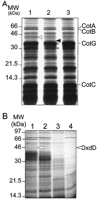
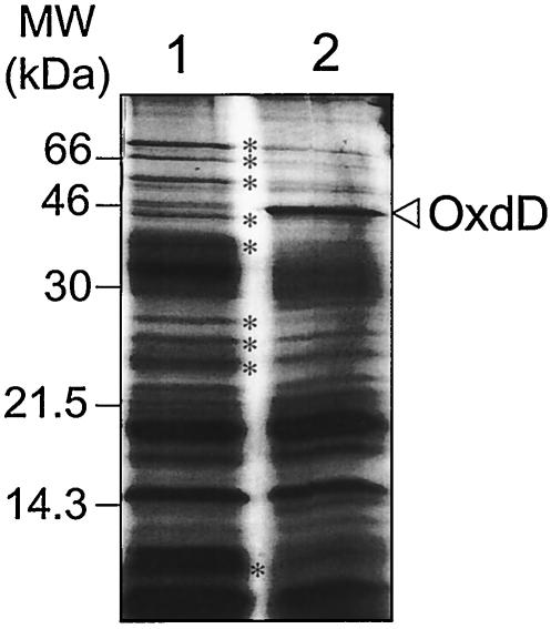
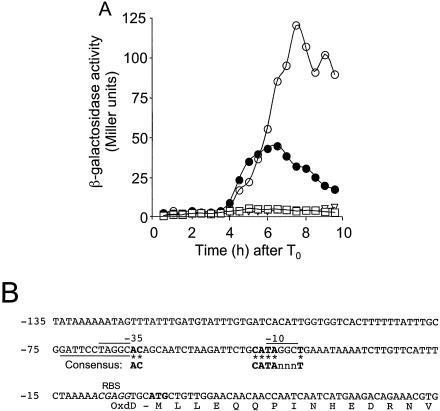
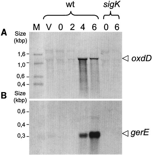
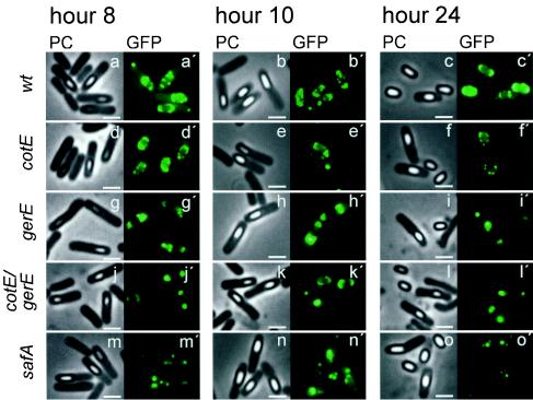
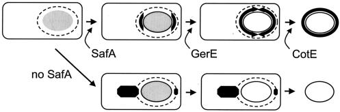
Over 30 polypeptides are synthesized at various times during sporulation in Bacillus subtilis, and they are assembled at the surface of the developing spore to form a multilayer protein structure called the coat. The coat consists of three main layers, an amorphous undercoat close to the underlying spore cortex peptidoglycan, a lamellar inner layer, and an electron-dense striated outer layer. The product of the B. subtilis oxdD gene was previously shown to have oxalate decarboxylase activity when it was produced in Escherichia coli and to be a spore constituent. In this study, we found that OxdD specifically associates with the spore coat structure, and in this paper we describe regulation of its synthesis and assembly. We found that transcription of oxdD is induced during sporulation as a monocistronic unit under the control of sigma(K) and is negatively regulated by GerE. We also found that localization of a functional OxdD-green fluorescent protein (GFP) at the surface of the developing spore depends on the SafA morphogenetic protein, which localizes at the interface between the spore cortex and coat layers. OxdD-GFP localizes around the developing spore in a cotE mutant, which does not assemble the spore outer coat layer, but it does not persist in spores produced by the mutant. Together, the data suggest that OxdD-GFP is targeted to the interior layers of the coat. Additionally, we found that expression of a multicopy allele of oxdD resulted in production of spores with increased levels of OxdD that were able to degrade oxalate but were sensitive to lysozyme.


Figures

FIG. 1.
FIG. 1.
Genetic organization of the oxdD locus of B. subtilis. The positions, lengths, and directions of transcription of the yoaO, oxdD (yoaN), and yoaM genes are indicated below a partial restriction map of the region (27). The stem-loop structures and the bent arrow preceding the oxdD gene indicate transcription terminators and the putative oxdD promoter, respectively. The inserts present in the plasmids are also indicated. All the plasmids are described in Materials and Methods.
FIG. 2.
FIG. 2.
Spore coat polypeptides extracted from spores of several strains. Spores were purified, and the coat proteins were extracted as described in Materials and Methods and electrophoretically resolved on SDS—15% PAGE gels. (A) Spore coat protein extracts of the following strains: MB24 (wild type) (lane 1), AH2898 (oxdD) (lane 2), and AH2873 (oxdD-gfp) (lane 3). (B) Profile of coat proteins extracted from spores of the following strains: MB24 (wild type) (lane 1), AH2898 (oxdD) (lane 2), AH2835 (cotE) (lane 3), and AH94 (gerE) (lane 4). The open and solid arrowheads indicate the positions of the OxdD and Hag proteins, respectively. The asterisks in panel A indicates the position of proteins that appear to be less abundant in the oxdD mutant. The positions of molecular mass markers (MW) are indicated on the left.
FIG. 3.
FIG. 3.
Analysis of a multicopy allele of oxdD. Spore coat proteins were extracted from purified spores as described in Materials and Methods and electrophoretically resolved on SDS—15% PAGE gels. Lane 1, AH2954 containing pMK3 (control plasmid); lane 2, AH2953 containing pTC149 (oxdDMC). The open arrowhead indicates the position of OxdD. The asterisks indicate the positions of polypeptides that are absent in strain AH2953 expressing a multicopy allele of oxdD or whose amounts are reduced. The positions of molecular mass markers (MW) are indicated on the left.
FIG. 4.
FIG. 4.
Regulation of oxdD-lacZ expression. (A) An oxdD-lacZ fusion was inserted at the amyE locus of various strains, and samples were taken at different times after the initiation of sporulation in DSM (T0) to assay for β-galactosidase accumulation. The following strains were used: AH2886 (amyE::oxdD-lacZ) (•), AH2890 (sigK::erm amyE::oxdD-lacZ) (□), and AH2891 (gerE36 amyE::oxdD-lacZ) (○). The endogenous levels of β-galactosidase production were determined in wild-type strain MB24 (▿). (B) Sequence of the putative oxdD promoter and the −10 and −35 sequences aligned with the consensus for σK-dependent promoters (16). Bases identical to the bases in the consensus sequence are indicated by boldface type and asterisks. n represents any base. The lines above the DNA sequence indicate bases in the putative −35 and −10 regions that match the bases in the core of the GerE binding site consensus sequence (TRGGY); the line below the sequence indicates a region in the complementary strand that matches the larger consensus region for GerE binding (RWWTRGGYnnY) (22). The ribosome binding site (RBS) is indicated by italics, and the start codon is indicated by boldface type just downstream of the ribosome binding site.
FIG. 5.
FIG. 5.
oxdD is monocistronic. A wild-type strain (wt) and a congenic sigK mutant were grown in DSM. Samples were taken during the exponential growth phase (vegetative growth [lane V]), at the onset of sporulation (time zero [lane 0]), and at various times throughout sporulation (times [in hours] are indicated after time zero [lanes 2, 4, and 6]). Total RNA was prepared as described in Materials and Methods. RNA samples were electrophoretically resolved on denaturing agarose-formaldehyde gels and transferred to nylon membranes. The RNA blots were hybridized with DIG-labeled probes complementary to the mRNA of oxdD (A) and gerE (B). Transcript sizes were determined based on the position of the DIG-labeled marker (lane M).
FIG. 6.
FIG. 6.
Assembly of OxdD-GFP into the spore coat. A functional OxdD-GFP fusion was introduced into a wild-type strain (wt) and into strains bearing mutations in loci known to be involved in assembly of the spore coat. The strains were grown in DSM, and samples were taken 8, 10, and 24 h after the onset of sporulation. Sporulating cells were observed by phase-contrast microscopy (PC) (a to o) and by fluorescence microscopy (a′ to o′) to detect OxdD-GFP (GFP). The following strains were used: AH2873 (oxdD-gfp), AH2883 (cotE oxdD-gfp), AH2912 (gerE oxdD-gfp), AH2913 (cotE gerE oxdD-gfp), and AH2944 (safA oxdD-gfp). Representative specimens are shown in each case. Quantification of the decoration patterns is shown in Table 4. Scale bars = 2 μm.
FIG. 7.
FIG. 7.
Model for the assembly of OxdD. The model predicts that OxdD is initially targeted to the polar cap regions of the developing spore in a safA-dependent manner. Localization of OxdD-GFP becomes apparent when the spore develops refractility (initially the spore is phase grey, as shown). In the absence of SafA, OxdD-GFP accumulates at the mother cell-prespore border as spots or patches, which persist until late times in development, but not in association with the released spore. Complete encircling of the developing spore by OxdD-GFP requires expression of gerE but not expression of cotE and occurs as the spore becomes phase bright (open ellipse). The requirement for safA and gerE but not for cotE suggests that OxdD associates with the inner coat layers. However, OxdD does not persist in stable association with the coat layers in the absence of the outer coat assembly in a cotE mutant. The dashed line indicates the position of the CotE ring, which marks the site of assembly of the outer coat. The CotE ring forms before the spore shows any signs of refractility or decoration by OxdD-GFP (initial cell).

Similar articles

Cited by

References

    1. Anand, R., P. C. Dorrestein, C. Kinsland, T. P. Begley, and S. E. Ealick. 2002. Structure of oxalate decarboxylase from Bacillus subtilis at 1.75 A resolution. Biochemistry 41:7659-7669. - PubMed
    1. Aronson, A. I., L. Ekanayake, and P. C. Fitz-James. 1992. Protein filaments may initiate the assembly of the Bacillus subtilis spore coat. Biochimie 74:661-667. - PubMed
    1. Aronson, A. I., H. Y. Song, and N. Bourne. 1989. Gene structure and precursor processing of a novel Bacillus subtilis spore coat protein. Mol. Microbiol. 3:437-444. - PubMed
    1. Bauer, T., S. Little, A. G. Stover, and A. Driks. 1999. Functional regions of the Bacillus subtilis spore coat morphogenetic protein CotE. J. Bacteriol. 181:7043-7051. - PMC - PubMed
    1. Beall, B., A. Driks, R. Losick, and C. P. Moran, Jr. 1993. Cloning and characterization of a gene required for assembly of the Bacillus subtilis spore coat. J. Bacteriol. 175:1705-1716. - PMC - PubMed

Publication types

MeSH terms

LinkOut - more resources