. 2004 Mar;186(5):1381-7.
doi: 10.1128/JB.186.5.1381-1387.2004.

Studies of the interaction of Escherichia coli YjeQ with the ribosome in vitro

Affiliations

Studies of the interaction of Escherichia coli YjeQ with the ribosome in vitro

Denis M Daigle et al. J Bacteriol. 2004 Mar.

Abstract

Escherichia coli YjeQ represents a conserved group of bacteria-specific nucleotide-binding proteins of unknown physiological function that have been shown to be essential to the growth of E. coli and Bacillus subtilis. The protein has previously been characterized as possessing a slow steady-state GTP hydrolysis activity (8 h(-1)) (D. M. Daigle, L. Rossi, A. M. Berghuis, L. Aravind, E. V. Koonin, and E. D. Brown, Biochemistry 41: 11109-11117, 2002). In the work reported here, YjeQ from E. coli was found to copurify with ribosomes from cell extracts. The copy number of the protein per cell was nevertheless low relative to the number of ribosomes (ratio of YjeQ copies to ribosomes, 1:200). In vitro, recombinant YjeQ protein interacted strongly with the 30S ribosomal subunit, and the stringency of that interaction, revealed with salt washes, was highest in the presence of the nonhydrolyzable GTP analog 5'-guanylylimidodiphosphate (GMP-PNP). Likewise, association with the 30S subunit resulted in a 160-fold stimulation of YjeQ GTPase activity, which reached a maximum with stoichiometric amounts of ribosomes. N-terminal truncation variants of YjeQ revealed that the predicted OB-fold region was essential for ribosome binding and GTPase stimulation, and they showed that an N-terminal peptide (amino acids 1 to 20 in YjeQ) was necessary for the GMP-PNP-dependent interaction of YjeQ with the 30S subunit. Taken together, these data indicate that the YjeQ protein participates in a guanine nucleotide-dependent interaction with the ribosome and implicate this conserved, essential GTPase as a novel factor in ribosome function.


Figures

FIG. 1.
FIG. 1.
Colocalization of YjeQ with ribosomes from E. coli MG1655 visualized by immunoblotting. Wild-type E. coli (4 liters) was grown in LB to an OD600 of 0.8, harvested by centrifugation at 8,500 × g for 15 min, and lysed by three consecutive passes through a French pressure cell at 10,000 lb/in2. (A) The lysate was clarified by centrifugation at 40,000 × g for 1 h, and both pellet (P) and supernatant (S) fractions were kept for analysis. (B) The supernatant was further clarified by ultracentrifugation at 150,000 × g for 2 h. (C through E) Subsequent washing and pelleting steps first with 0.5% Triton (C), then with 60 mM NH4Cl (D), and finally with 1 M NH4Cl (E) were performed by standard methods (13). At each step, pellets were resuspended in volumes identical to those of the supernatants for analysis. Immunoblotting employed SDS-15% polyacrylamide gels with a rabbit polyclonal antibody specific for YjeQ(21-350) as the primary antibody and HRP-conjugated donkey anti-rabbit IgG as the secondary antibody. Blots were developed by using the Western Lightning Chemiluminescence Reagent Plus kit (Perkin-Elmer, Boston, Mass.).
FIG. 2.
FIG. 2.
YjeQ variants constructed and purified in this study. (A) Scaled diagram showing the locations of motifs in YjeQ and the deletion variants constructed. (B) Five micrograms of the purified proteins was prepared by boiling in Laemmli buffer (14) containing 8% 2-mercaptoethanol prior to SDS-15% PAGE. The gels were visualized by staining with Coomassie brilliant blue R250. YjeQ variants characterized in this study are as follows: YjeQ(1-350) (39.1 kDa) (lane 1), YjeQ(21-350) (36.8 kDa) (lane 2), YjeQ(114-350) (27.9 kDa) (lane 3), and YjeQ(21-350) S221A (36.8 kDa) (lane 4).
FIG. 3.
FIG. 3.
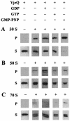
Binding of YjeQ to 70S ribosomes and ribosomal subunits revealed by immunoblotting. Full-length YjeQ(1-350) was tested for the ability to interact with 70S ribosomes and ribosomal subunits following a 1-h incubation in 20 mM Tris-HCl (pH 7.5)-10.5 mM magnesium acetate-60 mM NH4Cl-3 mM 2-mercaptoethanol at 30°C in the presence or absence of GDP, GTP, or GMP-PNP (2 mM). Reactions consisted of YjeQ and ribosomes, each at 2 μM. Samples (50 μl) were overlaid onto 20% (wt/vol) sucrose cushions (bed volume, 150 μl) and pelleted by ultracentrifugation at 513,000 × g in a Beckman Optima Max ultracentrifuge with a TLA 120.1 rotor for 1.5, 2, or 3 h for 70, 50, or 30S subunits, respectively. The pellets were resuspended in an equivalent volume (200 μl) of assay buffer, and supernatant (S) and pellet (P) fractions were mixed with 40 μl of sixfold-concentrated SDS-polyacrylamide gel electrophoresis loading buffer and separated by SDS-15% PAGE. Western blotting used a rabbit polyclonal antibody raised against YjeQ as the primary antibody and donkey anti-rabbit IgG coupled to HRP as the secondary antibody. (A) Binding of YjeQ to the 30S ribosomal subunit. (B) Binding of YjeQ to the 50S ribosomal subunit. (C) Binding of YjeQ to 70S ribosomes.
FIG. 4.
FIG. 4.
YjeQ binding to both the 30 and 50S ribosomal subunits in the presence of saturating levels of GMP-PNP (2 mM) analyzed by 10-to-30% (wt/vol) sucrose gradient ultracentrifugation. The reaction mixture consisted of 70S ribosomes (8 A260 units) purified by sucrose gradient ultracentrifugation (as described in Materials and Methods) and YjeQ, each at 3.7 μM. The sample (50 μl) was overlaid onto a 5-ml 10-to-30% (wt/vol) sucrose gradient and separated by ultracentrifugation at 43,000 × g in a Beckman Optima Max ultracentrifuge with an MLS 50 rotor for 16 h. (A) The gradient was fractionated as described in Materials and Methods, and fractions were analyzed by absorbance at 260 nm. (B) Selected fractions were separated by SDS-15% PAGE and analyzed by immunoblotting for YjeQ (as described in the legend to Fig. 3).
FIG. 5.
FIG. 5.
Salt stringency of the interaction of YjeQ(1-350) with the 30S ribosomal subunit. Immunoblotting of SDS-15% polyacrylamide gels separating pellet (P) and supernatant (S) fractions from the ribosomal pelleting assay (described in the legend to Fig. 3) was performed with increasing salt concentrations (KCl and NH4Cl) and with saturating (2 mM) levels of GMP-PNP or GDP as indicated.
FIG. 6.
FIG. 6.
Maximal stimulation of YjeQ GTPase at 1:1 stoichiometry with ribosomes. The GTPase activitiesof YjeQ and its variants were assessed by monitoring the steady-state release of phosphate from the enzyme by using the Malachite green-ammonium molybdate colorimetric assay described previously (9). All reactions were carried out at 30°C for 1 h, and reaction mixtures contained 200 nM YjeQ and saturating (2.5 mM) levels of GTP. The concentration of 30S ribosomes was varied from 3 to 800 nM. Sample data points are averages of duplicate reactions. The data were fit to a sigmoidal four-parameter equation by using SigmaPlot (version 8.0) to generate the curve shown.
FIG. 7.
FIG. 7.
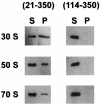
Ribosomal association by YjeQ N-terminal variants. The abilities of the YjeQ N-terminal deletion variants to bind various forms of the ribosome were assayed by a ribosomal pelleting assay (described in the legend to Fig. 3). Reaction components (50 μl) were separated following a 1-h incubation at 30°C of 100 pmol of YjeQ variant and 100 pmol of ribosome in 20 mM Tris-HCl (pH 7.5)-10.5 mM magnesium acetate-60 mM NH4Cl-3 mM 2-mercaptoethanol containing 2 mM GMP-PNP. Identical binding behavior was observed when assays contained 2 mM GDP instead of GMP-PNP (data not shown). The samples were overlaid onto 20% (wt/vol) sucrose cushions (bed volume, 150 μl) and pelleted by ultracentrifugation. The pellets (P) were resuspended in a volume (200 μl) equivalent to that of supernatants (S), and both fractions were separated by SDS-15% PAGE followed by immunoblot analysis (described in the legend to Fig. 3).

Similar articles

Cited by

References

    1. An, G., B. R. Glick, J. D. Friesen, and M. C. Ganoza. 1980. Identification and quantitation of elongation factor EF-P in Escherichia coli cell-free extracts. Can. J. Biochem. 97:23-28. - PubMed
    1. Aoki, H., K. Dekany, S.-L. Adams, and M. C. Ganoza. 1997. The gene encoding the elongation factor P protein is essential for viability and is required for protein synthesis. J. Biol. Chem. 272:32254-32259. - PubMed
    1. Arcus, V. 2002. OB-fold domains: a snapshot of the evolution of sequence, structure and function. Curr. Opin. Struct. Biol. 12:794-801. - PubMed
    1. Arigoni, F., F. Talabot, M. Peitsch, M. D. Edgerton, E. Meldrum, E. Allet, R. Fish, T. Jamotte, M. L. Curchod, and H. Loferer. 1998. A genome-based approach for the identification of essential bacterial genes. Nat. Biotechnol. 16:851-856. - PubMed
    1. Ausubel, F. M., R. Brent, R. E. Kingston, D. D. Moore, J. G. Seidman, J. A. Smith, and K. Struhl. 1994. Current protocols in molecular biology, vol. 2, chapter 11, section 11.12-11.13. John Wiley & Sons Inc., Boston, Mass.

MeSH terms

LinkOut - more resources