. 2004 Feb 15;64(4):1323-30.
doi: 10.1158/0008-5472.can-03-1816.

Imaging tri-fusion multimodality reporter gene expression in living subjects

Affiliations

Imaging tri-fusion multimodality reporter gene expression in living subjects

Pritha Ray et al. Cancer Res. .

Abstract

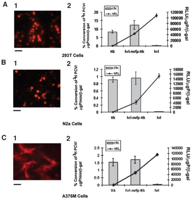
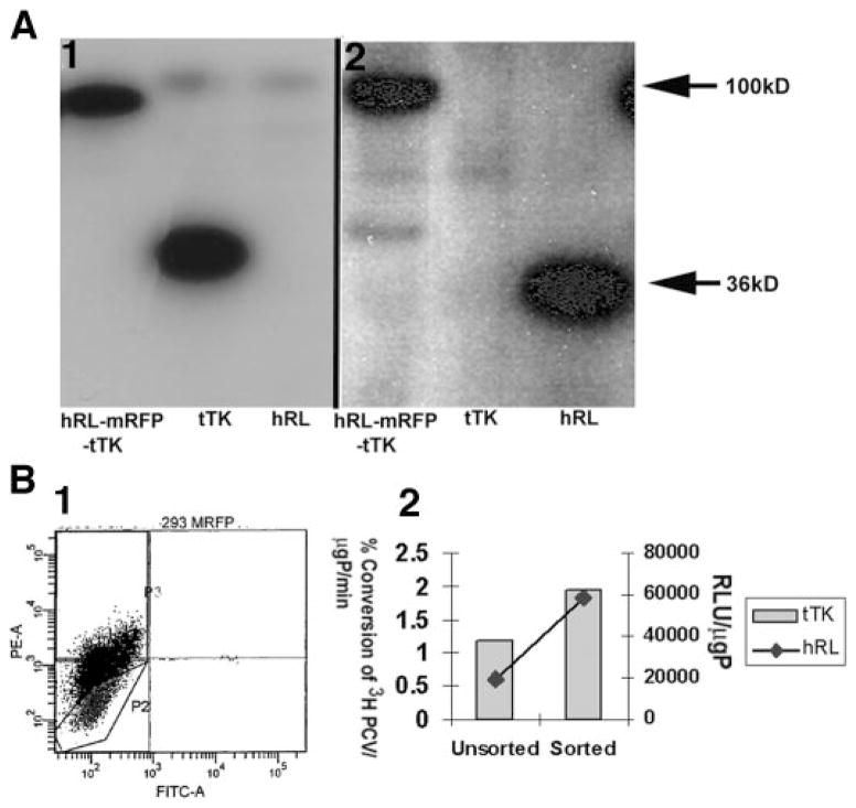
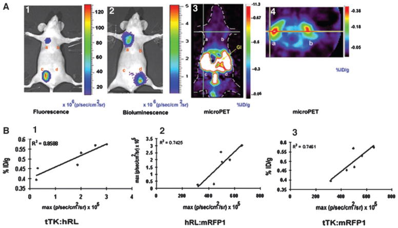
Imaging reporter gene expression in living subjects with various imaging modalities is a rapidly accelerating area of research. Applications of these technologies to cancer research, gene therapy, and transgenic models are rapidly expanding. We report construction and testing of several triple fusion reporter genes compatible with bioluminescence, fluorescence and positron emission tomography (PET) imaging. A triple fusion reporter vector harboring a bioluminescence synthetic Renilla luciferase (hrl) reporter gene, a reporter gene encoding the monomeric red fluorescence protein (mrfp1), and a mutant herpes simplex virus type 1 sr39 thymidine kinase [HSV1-truncated sr39tk (ttk); a PET reporter gene] was found to preserve the most activity for each protein component and was therefore investigated in detail. After validating the activities of all three proteins encoded by the fusion gene in cell culture, we imaged living mice bearing 293T cells transiently expressing the hrl-mrfp-ttk vector by microPET and using a highly sensitive cooled charge-coupled device camera compatible with both bioluminescence and fluorescence imaging. A lentiviral vector carrying the triple fusion reporter gene was constructed and used to isolate stable expressers by fluorescence-activated cell sorting. These stable 293T cells were further used to show good correlation (R(2) approximately 0.74-0.85) of signal from each component by imaging tumor xenografts in living mice with all three modalities. Furthermore, metastases of a human melanoma cell line (A375M) stably expressing the triple fusion were imaged by microPET and optical technologies over a 40-50-day time period in living mice. Imaging of reporter gene expression from single cells to living animals with the help of a single tri-fusion reporter gene will have the potential to accelerate translational cancer research.


Figures

Fig. 1
Fig. 1
mRFP1, hRL, and tTK activity exhibited by 293T, N2a, and A375M cell lines transiently transfected with the hrl-mrfp-ttk fusion construct. 293T (A), N2a (B), and A375M (C) cells were transiently cotransfected with CMV-β-gal and either CMV-hrl-mrfp-ttk, CMV-ttk, CMV-hrl, or CMV-mrfp1; harvested 24 h later; and assayed for mRFP1 expression (A.1, B.1, and C.1; by fluorescence microscopy) and tTK/hRL enzyme activities (A.2, B.2, and C.2). Bars for the fluorescence micrographs represent 100 μm. Values for tTK and hRL activity were normalized with β-galactosidase activity for each transfection. The tTK activity is expressed as (percentage of conversion of [8-3H]penciclovir to its phosphorylated form)/μg protein/min. The hRL activity is expressed as relative light units (RLU)/μg protein. Error bars represent SE for triplicate measurements. The slightly higher tTK activity exhibited by the hRL-mRFP-tTK fusion protein in comparison with the positive tTK protein is not statistically significant; however, the hRL activity of the hRL-mRFP-tTK fusion protein is significantly lower (P < 0.05) than the positive hRL protein.
Fig. 2
Fig. 2
Biochemical and flow cytometric characterization of hrl-mrfp-ttk fusion reporter gene expression. A, Western blot analysis of hRL-mRFP-tTK fusion protein. Twenty μg of total cellular protein obtained from the cell lysates of transiently transfected 293T cells with hrl-mrfp-ttk, ttk, and hrl plasmids were resolved in a 10% SDS poly-acrylamide gel and transferred and probed with anti-TK (A.1) and anti-RL (A.2) antibodies. A 100-kDa band was specifically recognized by the antibodies only from the hRL-mRFP-tTK fusion samples. The polyclonal anti-TK and the monoclonal anti-RL antibody recognize tTK (second lane in A.1) and hRL (third lane in A.2) at about 36 kDa, respectively. B, flow cytometry plot and enzymatic activities of the positive expressers of CS-hrl-mrfp-ttk (lentiviral vector)-infected 293T cells. One million 293T cells were infected with CS-hrl-mrfp-ttk vector and sorted with fluorescence-activated cell sorting with a filter at 585 ± 42 nm band setting. Highly fluorescing cells (~33%) that migrated to the P3 sector (B.1) were collected and further tested for tTK and hRL enzyme activities (B.2). The sorted fraction showed higher tTK and hRL activities than the unsorted population. The TK activity is expressed as (percentage of conversion of [8-3H]penciclovir to its phosphorylated form)/μg protein/min. The RL activity is expressed as relative light units (RLU)/μg protein.
Fig. 3
Fig. 3
Results of imaging living mice. A, fluorescence, bioluminescence, and micro-positron emission tomography (PET) imaging of hrl-mrfp-ttk expression in the same living nude mouse. Ten million 293T cells transiently expressing the CMV-hrl-mrfp-ttk, CMV-ttk, CMV-mrfp1, and CMV-hrl plasmids were implanted s.c. at four sites on the ventral side of a nude mouse and imaged the next day for fluorescence/bioluminescence and PET using a cooled charge-coupled device (CCD) camera and microPET, respectively. Fluorescence imaging was performed by placing the mouse in a CCD camera for 1 s, and a fluorescence image was acquired with a excitation filter at 500–550 nm and an emission filter at 575– 650 nm. Cells expressing the fusion (A.1, a) and mrfp1 (A.1, c) genes showed fluorescence, and the signal is recorded as maximum photons/sec/cm2/sr (A.1). The same mouse was then scanned in the CCD camera for bioluminescence after injection of coelenterazine via tail vein, and bioluminescence signal was found in cells expressing the fusion (A.2, a) and hrl (A.2, d) and recorded as maximum photons/sec/cm2/sr (A.2). After the optical scan, the same mouse was imaged by microPET using 9-(4-[18F]fluoro-3-hydroxymethylbutyl)guanine (FHBG). Cells expressing the fusion reporter gene (A.3, a and A.4, a) and ttk gene (A.3, b and A.4, b) showed FHBG accumulation in coronal section (A.3) and trans-axial section (A.4). Nonspecific accumulation of tracer was found in the gastrointestinal tracts and bladder (attributable to clearance of FHBG; A.3). B, in vivo correlation of hrl, mrfp1, and ttk gene expression exhibited by four clones of 293T cells stably but differentially expressing the hrl-mrfp-ttk fusion. Ten million cells of each clone were implanted on the axillary regions of the ventral side of three nude mice (two clones in each mouse), and after 24 h, mice were imaged by the cooled CCD camera and microPET. Plots of percentage of ID/g versus bioluminescence [expressed as maximum photons/second/centimeter2/steradian (p/s/cm2/sr); B.1], bioluminescence versus fluorescence (both expressed as maximum p/s/cm2/sr; B.2), and percentage of ID/g versus fluorescence (expressed as maximum p/s/cm2/sr; B.3) were obtained from the regions of interest drawn over the regions of cell implantation. Each of the six data points of each plot represents region of interest data from the fluorescence, bioluminescence, and microPET images of the same living mouse, with a total of three mice (2 points/mouse)
Fig. 4
Fig. 4
Multimodality imaging of metastasis of A375M cells stably expressing the hrl-mrfp-ttk fusion reporter gene in living mice. A, bioluminescence imaging of a SCID mouse injected with A375M cells expressing the hrl-mrfp-ttk vector at day 0. 7 × 10⁵ A375M cells stably expressing the triple fusion were injected via tail-vein in a SCID mouse and two hours later imaged for bioluminescence signal following tail-vein injection of coelenterazine. Prominent bioluminescence signal was found from the region of both the lungs [1.3–1.5 × 10⁵ max (p/sec/cm2/sr)]. B, bioluminescence imaging of the same SCID mouse at day 40. At day 40, the same mouse was imaged and relatively high bioluminescence signal [2 × 10⁵ max (p/sec/cm2/sr)] was found from the left lung region and moderate signal from the right lung region. A faint bioluminescence signal (5 × 10³ p/sec/cm2/sr) was also present from the right pelvic region. C, microPET imaging of the same SCID mouse at day 40. Following a bioluminescence scan, the mouse was imaged in microPET using FHBG. Shown is a thin coronal slice of ~1-mm thickness. A strong signal (~0.78% ID/g) was present from the chest region (Ch) with lower signal (0.35% ID/g) from the lung region. The stronger PET signal was found to be from a metastatic tumor present deep inside the body, as evident from the fluorescence photograph (4.E). Note the gallbladder (GB) retains FHBG so background signal from the GB is also seen in the microPET images. D, light photograph of the same SCID mouse after sacrifice and organ exposure (image has been modified by using Adobe Photoshop version 6). E, whole body fluorescence imaging of the same SCID mouse. Fluorescing metastatic tumors were found in lung and chest regions that correspond with the bioluminescence and PET images.

References

    1. Massoud T, Gambhir S. Molecular imaging in living subjects: seeing fundamental biological processes in a new light. Genes Dev. 2003;17:545–580. - PubMed
    1. Van Roessel P, Brand A. Imaging into the future: visualizing gene expression and protein interactions with fluorescent proteins. Nat Cell Biol. 2002;4:E15–E20. - PubMed
    1. Gambhir SS. Molecular imaging of cancer with positron emission tomography. Nat Rev Cancer. 2002;2:683–693. - PubMed
    1. Bremer C, Weissleder R. In vivo imaging of gene expression. Acad Radiol. 2001;8:15–23. - PubMed
    1. Vooijs M, Jonkers J, Lyons S, Berns A. Noninvasive imaging of spontaneous retinoblastoma pathway-dependent tumors in mice. Cancer Res. 2002;62:1862–1867. - PubMed

Publication types