. 2004 Mar;16(3):672-93.
doi: 10.1105/tpc.019703. Epub 2004 Feb 18.

Identification of multivesicular bodies as prevacuolar compartments in Nicotiana tabacum BY-2 cells

Affiliations

Identification of multivesicular bodies as prevacuolar compartments in Nicotiana tabacum BY-2 cells

Yu Chung Tse et al. Plant Cell. 2004 Mar.

Abstract

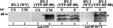
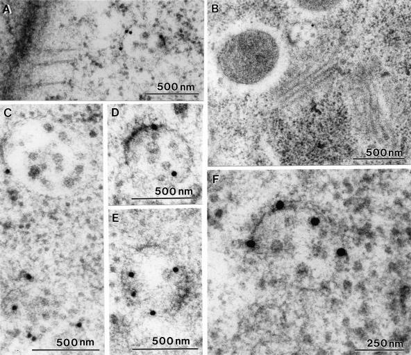
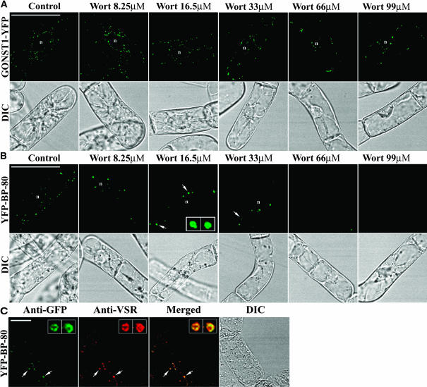
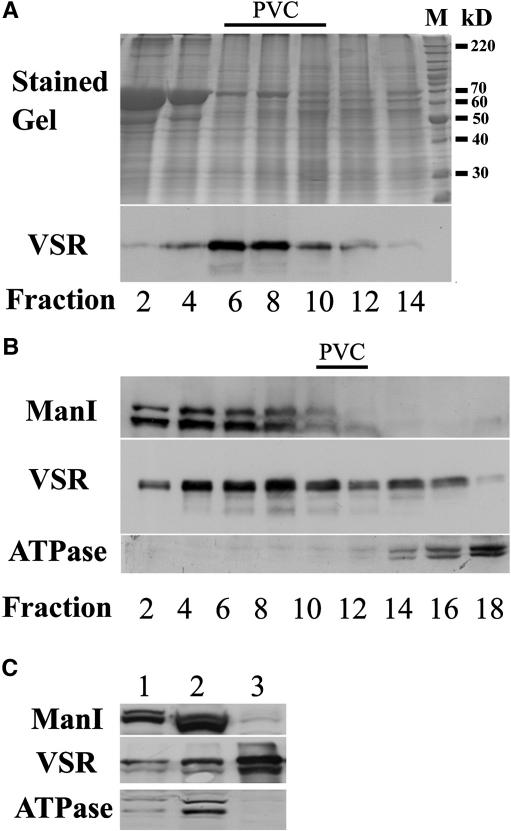
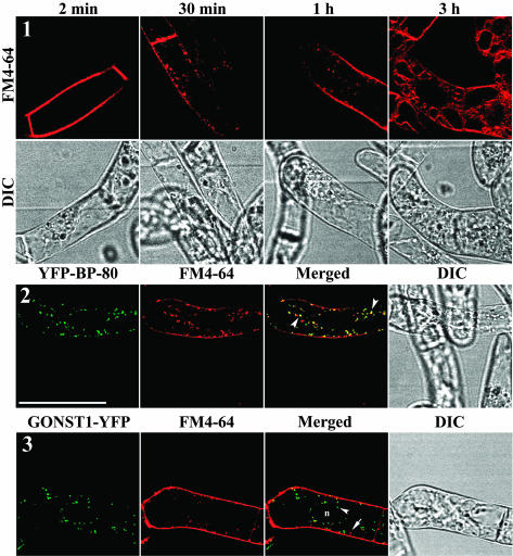
Little is known about the dynamics and molecular components of plant prevacuolar compartments (PVCs). We have demonstrated recently that vacuolar sorting receptor (VSR) proteins are concentrated on PVCs. In this study, we generated transgenic Nicotiana tabacum (tobacco) BY-2 cell lines expressing two yellow fluorescent protein (YFP)-fusion reporters that mark PVC and Golgi organelles. Both transgenic cell lines exhibited typical punctate YFP signals corresponding to distinct PVC and Golgi organelles because the PVC reporter colocalized with VSR proteins, whereas the Golgi marker colocalized with mannosidase I in confocal immunofluorescence. Brefeldin A induced the YFP-labeled Golgi stacks but not the YFP-marked PVCs to form typical enlarged structures. By contrast, wortmannin caused YFP-labeled PVCs but not YFP-labeled Golgi stacks to vacuolate. VSR antibodies labeled multivesicular bodies (MVBs) on thin sections prepared from high-pressure frozen/freeze substituted samples, and the enlarged PVCs also were indentified as MVBs. MVBs were further purified from BY-2 cells and found to contain VSR proteins via immunogold negative staining. Similar to YFP-labeled Golgi stacks, YFP-labeled PVCs are mobile organelles in BY-2 cells. Thus, we have unequivocally identified MVBs as PVCs in N. tabacum BY-2 cells. Uptake studies with the styryl dye FM4-64 strongly indicate that PVCs also lie on the endocytic pathway of BY-2 cells.


Figures

Figure 1.
Figure 1.
Characterization of VSR and BP-80 CT Antibodies. (A) Anti-VSR detection of VSR proteins in Arabidopsis (Arab), P. sativum (pea), and BY-2 cells from total protein extraction. (B) and (C) Anti-VSR and anti-BP-80 CT detection of VSR proteins from soluble (CS) and membrane (CM) fractions in BY-2 cells and Arabidopsis, respectively. M, molecular marker in kilodaltons.
Figure 2.
Figure 2.
Colocalization of VSR Antibodies with PVC Markers. Shown are double-labeling of VSR antibodies with other known PVC markers, AtSYP21 and 14G7, in fixed P. sativum root tip cells. The yellow appearance in the merged images indicates colocalization of two antibodies. Bar = 10 μm.
Figure 3.
Figure 3.
Development of Transgenic BY-2 Cell Lines Expressing the YFP-BP-80 and GONST1-YFP Reporters. Confocal images collected from living cells expressing the Golgi marker GONST1-YFP (panel 1) and the PVC marker YFP-BP-80 (panel 2), showing similar punctate patterns. Panels 3 and 4 showed colocalization between YFP signals derived either from YFP reporters (green) or anti-GFP signals (red) in fixed cells. Colocalization of two signals was indicated by a yellow color. Arrowheads and arrows indicate colocalization (panels 3 to 4) of two signals. n, nucleus; DIC, differential interference contrast. Bar = 50 μm.
Figure 4.
Figure 4.
Protein Gel Blot Analysis of Transgenic BY-2 Cells Expressing the YFP-BP-80 Reporter. Lanes 3 to 6, anti-GFP detection of the full-length YFP-BP-80 reporter proteins in membrane (CM) fraction (lane 4, asterisk) and vacuole fraction (V) (lane 5, asterisk); lane 8, anti-BP-80 CT detection of the YFP-BP-80 reporter (asterisk) in vacuole fraction. The tentative degradation products derived from the reporter proteins are indicated by double asterisks (lanes 5 and 6). Wild-type proteins also were included as controls (lanes 1 and 2). CS, soluble fraction; P, pellet.
Figure 5.
Figure 5.
Anti-VSR Colocalizes with the YFP-BP-80 Reporter but Is Separate from the GONST1-YFP Reporter. Transgenic N. tabacum BY-2 cells expressing either YFP-BP-80 or GONST1-YFP reporters were fixed and incubated with either VSR, AtSRY21, or ManI antibodies to detect PVC and Golgi stacks, respectively. The primary antibodies were detected with rhodamine-conjugated secondary antibodies (red), whereas the two YFP reporters (green) were ready for detection in the confocal microscope using a 488-nm laser. When green and red images were superimposed, colocalization of two signals is indicated by a yellow color. Arrowheads and arrows indicate colocalization and separation, respectively. n, nucleus. Bar = 50 μm.
Figure 6.
Figure 6.
Immunogold Labeling of MVBs with VSR Antibodies. (A) to (F) Thin sections prepared from high-pressure frozen/freeze-substituted samples were stained with VSR antibodies. Various MVBs are depicted that are labeled specifically with VSR antibodies.
Figure 7.
Figure 7.
BFA at 10 μg/mL Induces Structural Changes in YFP-Marked Golgi Stacks but Not in YFP-Marked PVCs. Transgenic BY-2 cells expressing either the GONST1-YFP reporter (A) or the YFP-BP-80 reporter (B) were incubated with BFA at 10 μg/mL for up to 1 h. Treated cells then were collected at the times indicated for YFP imaging in the confocal laser scanning microscope. Arrows indicate BFA-induced aggregates in cells expressing the GONST1-YFP Golgi marker. n, nucleus. Bars = 50 μm.
Figure 8.
Figure 8.
Wortmannin Induces the YFP-BP-80–Labeled PVCs to Form Small Vacuoles in a Dose-Dependent Manner. (A) and (B) Transgenic cells expressing the Golgi (A) and PVC (B) reporters were incubated with wortmannin (Wort) at various concentrations as indicated for 3 h before the treated cells were sampled for YFP imaging. Arrows in (B) indicate small vacuoles derived directly from the YFP-BP-80–labeled PVCs. The insets in the third panel of (B) are enlarged vacuole images of the two structures indicated by the arrows. (C) Colocalization of anti-VSR (red) with PVC-derived vacuoles in transgenic cells expressing the YFP-BP-80 reporter after treatment with wortmannin at 16.5 μM for 3 h. The insets are enlarged vacuole images of the two structures indicated by the arrows. n, nucleus. Bar in (A) and (B) = 50 μm; bar in (C) = 20 μm.
Figure 9.
Figure 9.
Time Course of Wortmannin-Induced PVC Vacuole Formation. (A) and (B) Transgenic BY-2 cell lines expressing the Golgi and PVC reporters were incubated with wortmannin at 33 μM for the times indicated. Samples of treated cells then were taken for YFP imaging. Arrowheads in (B) indicate wortmannin-induced vacuole formation. Insets are vacuolated YFP-BP-80–labeled PVCs. n, nucleus. Bars = 50 μm.
Figure 10.
Figure 10.
Ultrastructural Analysis of BFA on GONST1-YFP–Transformed BY-2 Cells. (A) Golgi stack from an untreated cell. c, cis face; t, trans face. (B) ER-Golgi hybrid. Note the outermost Golgi cisternae at both faces have an ER-like structure (arrowheads). (C) and (D) Cup-shaped Golgi structures sectioned in different planes. (E) Multiple Golgi stack aggregates adjacent to a multivesicular body (arrow) that has remained unchanged in size and morphology (Figure 11).
Figure 11.
Figure 11.
Ultrastructural Analysis of Wortmannin Effects on BY-2 Cells. (A) Golgi stack in an untreated cell. c, cis-cisterna; t, trans-cisterna. (B) Size comparison of Golgi stacks and MVBs (arrows) in an untreated cell. (C) High magnification of a multivesicular body in an untreated cell. (D) and (E) Highly stainable plaques (arrowheads) at the cytoplasmic surface of MVBs in untreated cells. (F) Golgi stack in a cell treated with wortmannin for 1 h. Stack parameters (number of cisternae, polarity) appear unchanged. (G) and (H) MVBs (chevrons) swell and show reduced numbers of internal vesicles in wortmannin-treated cells.
Figure 12.
Figure 12.
Isolation of PVCs from BY-2 Cells. (A) Identification of fractions enriched in VSR proteins. Cell homogenates were layered onto a discontinuous sucrose density gradient (20 and 60% [w/w]). Fractions were collected and subjected to SDS-PAGE followed by immunodetection with VSR antibodies. M, molecular marker in kilodaltons. (B) The VSR-eniched fractions from (A) (fractions 6 to 8) were further layered onto a continuous sucrose density gradient (25 to 50% [w/w]) and centrifuged isopycnically. Fractions were collected for protein gel blotting using various antibodies. (C) Progressive enrichment of VSRs in subcellular fractionation. Total protein from BY-2 cell homogenate (lane 1), VSR-enriched fractions 6 to 8 of (A) (lane 2), and fraction 11 from (B) (lane 3) were used for protein gel blot analysis using various antibodies. Equal amounts of protein (15 μg) applied to each lane.
Figure 13.
Figure 13.
Immunogold Negative Staining of Isolated PVCs. (A) Morphology of freshly isolated PVCs in VSR-enriched fraction 11 from Figure 12. (B) to (E) Various PVCs after immunogold staining with VSR antibodies. (F) and (G) Control labeling of isolated PVCs using α-TIP and secondary antibodies alone, respectively. Bars = 200 nm.
Figure 14.
Figure 14.
Colocalization of YFP-BP-80–Labeled PVCs with Internalized FM4-64–Marked Organelles in BY-2 Cells. Panel 1, uptake process of FM4-64 in living cells. Shown are steps of FM4-64 uptake profiled in living BY-2 cells in which the dye is first detected on the plasma membrane, followed by localization in internalized endosome-like structures and eventually insertion in the tonoplast. Panels 2 and 3, colocalization of PVC reporters with internalized FM4-64–labeled organelles. At 30 min after uptake, FM4-64–marked organelles colocalized with YFP-labeled PVCs in cells expressing the YFP-BP-80 reporter (panel 2) but remained separate from YFP-labeled Golgi stacks in cells expressing the GONST1-YFP reporter (panel 3). Bar = 50 μm.

References

    1. Ahmed, S.U., Bar-Peled, M., and Raikhel, N.V. (1997). Cloning and subcellular location of an Arabidopsis receptor-like protein that shares common features with protein-sorting receptors of eukaryotic cells. Plant Physiol. 114, 325–336. - PMC - PubMed
    1. Ahmed, S.U., Rojo, E., Kovaleva, V., Venkataraman, S., Dombrowski, J.E., Matsuoka, K., and Raikhel, N.V. (2000). The plant vacuolar sorting receptor AtELP is involved in transport of NH(2)-terminal propeptide-containing vacuolar proteins in Arabidopsis thaliana. J. Cell Biol. 149, 1335–1344. - PMC - PubMed
    1. Aniento, F., Helms, J.B., and Memon, A.R. (2003). How to make a vesicle: Coat protein-membrane interactions. Annu. Plant Rev. 9, 36–62.
    1. Baldwin, T.C., Handford, M.G., Yuseff, M.I., Orellana, A., and Dupree, P. (2001). Identification and characterization of GONST1, a golgi-localized GDP-mannose transporter in Arabidopsis. Plant Cell 13, 2283–2295. - PMC - PubMed
    1. Barr, F.A. (2002). Inheritance of the endoplasmic reticulum and Golgi apparatus. Curr. Opin. Cell Biol. 14, 496–499. - PubMed

Publication types

MeSH terms

LinkOut - more resources