. 2004 Mar;16(3):694-708.
doi: 10.1105/tpc.017400. Epub 2004 Feb 18.

Protein phosphorylation in amyloplasts regulates starch branching enzyme activity and protein-protein interactions

Affiliations

Protein phosphorylation in amyloplasts regulates starch branching enzyme activity and protein-protein interactions

Ian J Tetlow et al. Plant Cell. 2004 Mar.

Abstract

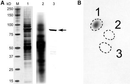
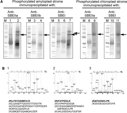
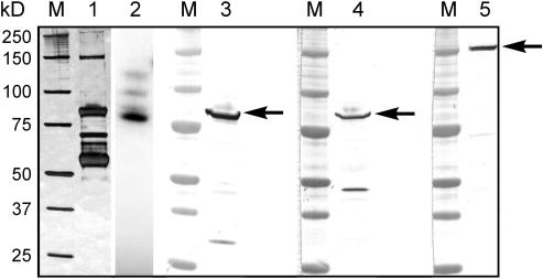
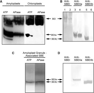
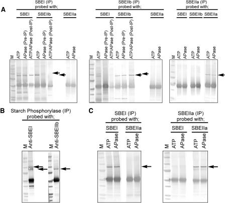
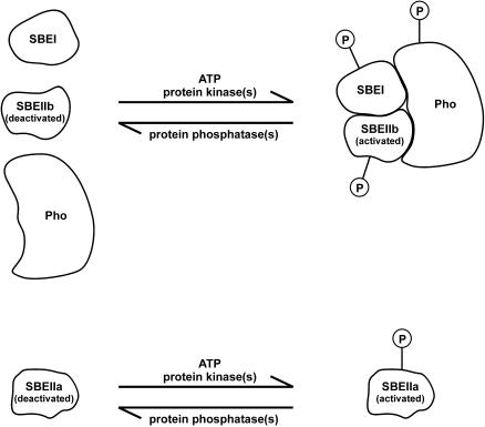
Protein phosphorylation in amyloplasts and chloroplasts of Triticum aestivum (wheat) was investigated after the incubation of intact plastids with gamma-(32)P-ATP. Among the soluble phosphoproteins detected in plastids, three forms of starch branching enzyme (SBE) were phosphorylated in amyloplasts (SBEI, SBEIIa, and SBEIIb), and both forms of SBE in chloroplasts (SBEI and SBEIIa) were shown to be phosphorylated after sequencing of the immunoprecipitated (32)P-labeled phosphoproteins using quadrupole-orthogonal acceleration time of flight mass spectrometry. Phosphoamino acid analysis of the phosphorylated SBE forms indicated that the proteins are all phosphorylated on Ser residues. Analysis of starch granule-associated phosphoproteins after incubation of intact amyloplasts with gamma-(32)P-ATP indicated that the granule-associated forms of SBEII and two granule-associated forms of starch synthase (SS) are phosphorylated, including SSIIa. Measurement of SBE activity in amyloplasts and chloroplasts showed that phosphorylation activated SBEIIa (and SBEIIb in amyloplasts), whereas dephosphorylation using alkaline phosphatase reduced the catalytic activity of both enzymes. Phosphorylation and dephosphorylation had no effect on the measurable activity of SBEI in amyloplasts and chloroplasts, and the activities of both granule-bound forms of SBEII in amyloplasts were unaffected by dephosphorylation. Immunoprecipitation experiments using peptide-specific anti-SBE antibodies showed that SBEIIb and starch phosphorylase each coimmunoprecipitated with SBEI in a phosphorylation-dependent manner, suggesting that these enzymes may form protein complexes within the amyloplast in vivo. Conversely, dephosphorylation of immunoprecipitated protein complex led to its disassembly. This article reports direct evidence that enzymes of starch metabolism (amylopectin synthesis) are regulated by protein phosphorylation and indicate a wider role for protein phosphorylation and protein-protein interactions in the control of starch anabolism and catabolism.


Figures

Figure 1.
Figure 1.
Phosphorylation of Amyloplast Stromal Proteins from T. aestivum Endosperm. Intact amyloplasts were isolated from developing T. aestivum endosperm at between 12 and 25 DAP and incubated with 100 μM γ-32P-ATP for 15 min at 25°C. Reactions were terminated, organelles lysed, and stromal phosphoproteins partially purified by affinity chromatography as described in Methods. (A) Eluted phosphoproteins were separated by 1D-SDS-PAGE. Lane 1, silver-stained gel of stromal phosphoproteins eluted from affinity column. Lane 2, an autoradiograph of the 32P-labeled stromal phosphoproteins. Lane 3, immunoblot of the protein fraction from lane 1 probed with anti-E. coli branching enzyme antibodies. M, molecular mass markers whose individual sizes are shown at the left in kilodaltons; the arrow at the right indicates the position of the 87- to 88-kD polypeptides (putative SBEs) that were excised for phosphoamino acid analysis. (B) Autoradiograph of phosphoamino acids separated by 2D thin-layer electrophoresis after acid hydrolysis of the 32P-labeled 87- to 88-kD polypeptide(s) shown in (A). The marked zones are positions of standards: 1, phosphoserine; 2, phosphothreonine; and 3, phosphotyrosine.
Figure 2.
Figure 2.
Identification of the Phosphorylated 87- to 88-kD Polypeptides from Plastids as SBE Isoforms. Intact amyloplasts (isolated from T. aestivum endosperm 12 to 25 DAP, 1.2 to 1.5 mg protein cm−3) and chloroplasts (isolated from 8- to 10-d-old T. aestivum leaves, 1.4 to 1.8 mg protein cm−3) were incubated with 100 μM γ-32P-ATP for 20 min at 25°C. Reactions were terminated, organelles lysed, and the stromal phosphoproteins partially purified by affinity chromatography as described in Methods. (A) Stromal phosphoproteins were incubated with different, peptide-specific, anti-SBE antisera, and the 87- to 88-kD phosphoproteins were immunoprecipitated as described in Methods. Lanes 1, 3, 5, 8, and 10, immunoblots of the immunoprecipitated phosphoproteins probed with the respective anti-SBE antisera. Lane 6, an immunoblot of amyloplast stromal proteins immunoprecipitated with anti-SBEI antibodies and probed with anti-H. vulgare endosperm (plastidial) starch phosphorylase antbodies. Lanes 2, 4, 7, 9, and 11, autoradiographs of the respective immunoblots. M, molecular mass markers. Arrows indicate the positions of the phosphorylated 87- to 88-kD polypeptides that cross-reacted with the respective anti-SBE antibodies. (B) Q-TOF-MS data of peptides from the 87- to 88-kD phosphoproteins immunoprecipitated with different anti-SBE antibodies and separated by 2D-PAGE. Panels 1 to 3, the MS survey acquisition data obtained for the 87- to 88-kD phosphoproteins from amyloplast stroma immunoprecipitated with the following: 1, anti-SBEIIa antibodies; 2, anti-SBEIIb antibodies; and 3, anti-SBEI antibodies. The data presented are for single representative analyses and in each case show the spectra obtained for one of the peptides. Below them are the corresponding sequences from each spectrum (in bold) and the sequences of other peptides acquired from the same sample.
Figure 3.
Figure 3.
Analysis and Identification of Granule-Associated Phosphoproteins from T. aestivum Endosperm Amyloplasts. Intact amyloplasts were isolated from developing endosperm at 12 to 25 DAP and incubated with 100 μM γ-32P-ATP for 20 min at 25°C. Reactions were terminated and organelles lysed and fractionated as described in Methods. Granule-associated proteins were extracted from starch grains after incubation of amyloplasts with γ-32P-ATP and separated by 1D-SDS-PAGE on 4 to 12% gradient gels. Lane 1, silver-stained polyacrylamide gel of polypeptides extracted from starch granules (15 μg of protein per lane); lane 2, autoradiograph of starch granule–associated polypeptides separated by SDS-PAGE (from lane 1), showing phosphorylated polypeptides; lanes 3 to 5, immunoblots of radiolabeled starch granule–associated polypeptides probed with peptide-specific anti-SBE-antibodies: lane 3, anti-SBEIIa; lane 4, anti-SBEIIb; and lane 5, anti-SBEI. M, molecular mass markers. Arrows indicate the positions of the starch granule–associated polypeptides that cross-reacted with the respective anti-SBE antibodies.
Figure 4.
Figure 4.
Activity Gel Analysis of SBE Isoforms Showing Effects of Dephosphorylation on Enzyme Activity. (A) Zymogram analysis of SBE activity from stromal extracts of amyloplasts (isolated from endosperm 12 to 25 DAP) and chloroplasts (from 8- to 10-d-old leaves) after pretreatment of the samples with 1 mM ATP or 10 units APase for 20 min at 25°C. Approximately 80 μg of amyloplast stromal protein per lane and 120 μg of chloroplast stromal protein per lane were separated on a 7-cm native polyacrylamide gel containing substrates for SBE. SBE activities were visualized by staining with I2/KI. (B) Immunoblots from SBE zymogram gels of amyloplast and chloroplast stromal proteins (same protein loadings as in [A]) developed with antisera against SBEI (lanes 1 and 2), SBEIIa (lanes 3 and 4), and SBEIIb (lanes 5 and 6), showing the positions of each of the different SBE forms on the zymogram. The antisera used in the immunoblots in Figure 4B were used at the same dilutions as for the immunoprecipitation experiments. Lanes 1, 3, and 5 contain amyloplast proteins, and lanes 2, 4, and 6 contain chloroplast proteins. (C) Zymogram analysis of granule-associated SBE activity in amyloplasts. Starch granule–associated proteins were extracted after incubation of plastids with ATP and subsequently either untreated or incubated with APase (see above). Samples were separated on native gels (∼80 μg of protein per lane) and SBE activity visualized as above. (D) Immunoblots from SBE zymogram gels of granule-associated proteins from amyloplasts (ATP treated) developed with various anti-SBE antisera as shown.
Figure 5.
Figure 5.
Coimmunoprecipitation of Plastid Stromal Proteins with Peptide-Specific Anti-SBE Antibodies. (A) Amyloplast lysates (1.2 to 1.5 mg protein cm−3) were either preincubated with 1 mM ATP or with 10 units of APase (Pre-IP) for 20 min at 25°C. Alternatively, amyloplast lysates were preincubated with 1 mM ATP and immunoprecipitated, and the washed immunoprecipitation products treated with 10 units of alkaline phosphatase (ATP/APase [Post-IP]). After immunoprecipitation (IP) with anti-SBE antibodies, immunoprecipitated proteins were separated by 1D-SDS-PAGE, electroblotted onto nitrocellulose, and developed with various anti-SBE antisera as shown. Bottom arrow indicates positions of 87- to 88-kD SBE proteins cross-reacting with anti-SBE antisera. Top arrow indicates a 110-kD stromal phosphoprotein (identified as starch phosphorylase) that cross-reacted with anti-SBEI antibodies and coimmunoprecipitated with phosphorylated SBEI. M, molecular mass markers. (B) Amyloplast lysates (1.2 to 1.5 mg protein cm−3) were preincubated with ATP (as above) and immunoprecipitated with anti-starch phosphorylase antibodies, separated by 1D-SDS-PAGE, immunoblotted, and developed with anti-SBEI or anti-SBEIIb antisera. (C) Chloroplast lysates (1.4 to 1.6 mg protein cm−3) made from plastids harvested at the end of the photoperiod were preincubated with either ATP or APase, immunoprecipitated with anti-SBE antibodies, separated by 1D-SDS-PAGE, immunoblotted, and developed with anti-SBEI and anti-SBEIIa antisera as described for amyloplasts in (A) and (B).
Figure 6.
Figure 6.
Model of Phosphorylation-Dependent Protein Complex Formation Involved in Starch Synthesis. Activation of SBEIIa and activation and complex formation involving SBEI, SBEIIb, and starch phosphorylase (Pho) by protein phosphorylation in the amyloplast stroma stimulate amylopectin biosynthesis. The functional relationships between the different components of the putative protein complex are unclear.

Similar articles

Cited by

References

    1. Altschul, S.F., Madden, T.L., Schäffer, A., Zhang, J., Zhang, Z., Miller, W., and Lipman, D.J. (1997). Gapped BLAST and PSI-BLAST: A new generation of protein database search programs. Nucleic Acids Res. 25, 3389–3402. - PMC - PubMed
    1. Båga, M., Nair, R.B., Repellin, A., Scoles, G.J., and Chibbar, R.N. (2000). Isolation of a cDNA encoding a granule-bound 152-kilodalton starch-branching enzyme in wheat. Plant Physiol. 124, 253–263. - PMC - PubMed
    1. Ball, S., Guan, H.-P., James, M., Myers, A., Keeling, P., Mouille, G., Buléon, A., Colonna, P., and Preiss, J. (1996). From glycogen to amylopectin: A model for the biogenesis of the plant starch granule. Cell 86, 349–352. - PubMed
    1. Ball, S.G., and Morell, M.K. (2003). From bacterial glycogen to starch: Understanding the biogenesis of the plant starch granule. Annu. Rev. Plant Biol. 54, 207–233. - PubMed
    1. Beatty, M.K., Rahman, A., Cao, H., Woodman, W., Lee, M., Myers, A.M., and James, M.G. (1999). Purification and molecular genetic characterization of ZPU1, a pullulanase-type starch-debranching enzyme from maize. Plant Physiol. 119, 255–266. - PMC - PubMed

Publication types

Substances

Associated data

LinkOut - more resources