. 2004 Feb 18;32(3):e33.
doi: 10.1093/nar/gnh029.

Surfection: a new platform for transfected cell arrays

Affiliations

Surfection: a new platform for transfected cell arrays

Fu-Hsiung Chang et al. Nucleic Acids Res. .

Abstract

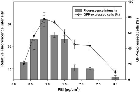
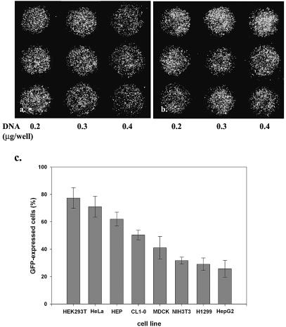
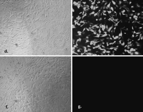
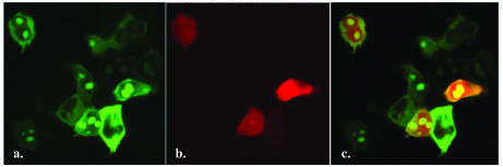
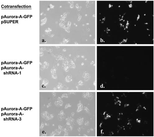
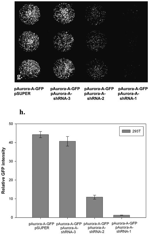
Efficient high-throughput expression of genes in mammalian cells can facilitate large-scale functional genomic studies. Towards this aim, we developed a simple yet powerful method to deliver genes into cells by cationic polymers on the surface of substrates. Transfection can be achieved by directly contacting nucleic acid-cell mixtures with the cationic substrates, e.g. polyethylenimine/collagen-coated wells. This single-step matrix-surface- mediated transfection method, termed 'surfection', can efficiently deliver multiple plasmids into cells and can successfully assay siRNA-mediated gene silencing. This technology represents the easiest method to transfer combinations of genes in large-scale arrays, and is a versatile tool for live-cell imaging and cell-based drug screening.


Figures

Figure 1
Figure 1
Effect of PEI concentration on transfection efficiency in 293T cells. The mini-wells (3 mm in diameter) were coated with mixtures of PEI (0.3– 3.0 µg/cm2) and gelatin (1 µg/cm2). Six thousand 293T cells in culture medium containing 10% fetal calf serum and pEGFP plasmid DNA (0.3 µg/well) were aliquoted directly into each well. The fluorescent image was taken 40 h after seeding the cells. Data are expressed as a percentage of GFP-expressing cells in the total cell population, and relative fluorescent intensity using MetaMorph software (mean ± standard deviation, obtained from triplicate wells) with luciferase DNA (pLuc) transfected cells as the background control. The experiment was repeated three times with similar results.
Figure 2
Figure 2
Genes delivered into cultured cells in polymer-coated wells. The mini-wells (3 mm in diameter) were coated with a mixture of PEI (1 µg/cm2) and gelatin (1 µg/cm2). Cells (2000–8000) in culture medium containing 10% fetal calf serum and plasmid DNA were aliquoted directly into each well. The fluorescent image was taken 40 h after seeding the cells. The dose effect of pEGFP DNA (0.2, 0.3 and 0.4 µg/well, respectively) on the transfection of (a) HeLa and (b) 293T cells is shown in triplicate. Magnification 40×. (c) The relative transfection efficiency of various cell lines was quantified by counting GFP-expressed cells in the total cell population (mean ± standard deviation, obtained from triplicate wells). A similar experiment was performed in a 96-well plate coated with PEI/RGD-PEI/collagen (1.8, 0.18 and 0.3 µg/cm2, individually) mixture using HeLa cells (2 × 10⁴) and 1 µg of pEGFP (d and e) or pLuc (f and g). The fluorescent image was taken 24 h after seeding the cells. Magnification 100×. The experiments were repeated three times with similar results.
Figure 2
Figure 2
Genes delivered into cultured cells in polymer-coated wells. The mini-wells (3 mm in diameter) were coated with a mixture of PEI (1 µg/cm2) and gelatin (1 µg/cm2). Cells (2000–8000) in culture medium containing 10% fetal calf serum and plasmid DNA were aliquoted directly into each well. The fluorescent image was taken 40 h after seeding the cells. The dose effect of pEGFP DNA (0.2, 0.3 and 0.4 µg/well, respectively) on the transfection of (a) HeLa and (b) 293T cells is shown in triplicate. Magnification 40×. (c) The relative transfection efficiency of various cell lines was quantified by counting GFP-expressed cells in the total cell population (mean ± standard deviation, obtained from triplicate wells). A similar experiment was performed in a 96-well plate coated with PEI/RGD-PEI/collagen (1.8, 0.18 and 0.3 µg/cm2, individually) mixture using HeLa cells (2 × 10⁴) and 1 µg of pEGFP (d and e) or pLuc (f and g). The fluorescent image was taken 24 h after seeding the cells. Magnification 100×. The experiments were repeated three times with similar results.
Figure 3
Figure 3
High efficiency of multiple gene transfer in cultured cells. Five thousand 293T cells in culture medium containing 10% fetal calf serum and three plasmid DNAs (0.1 µg/well, individually) were aliquoted directly into the PEI/gelatin-coated wells (3 mm in diameter). The fluorescent image was taken 40 h after seeding the cells. Panels show the subcellular location of CRMP-1 protein fused with GFP (a, cytoplasm), NoBP protein fused with GFP (a, nucleoli), and DsRed fluorescent protein (b) in human 293T cells after triple gene transfer. (c) is the merged picture of (a) and (b). Magnification 400×. The cells were 80% confluent when the image was recorded.
Figure 4
Figure 4
shRNA-mediated gene silencing. Five thousand 293T cells in culture medium containing 10% fetal calf serum and plasmid DNA were aliquoted directly into the PEI/gelatin-coated wells (3 mm in diameter). These cells were cotransfected with equal amounts (0.15 µg of each plasmid per well) of reporter plasmid containing Aurora-A gene fused to GFP (pAurora-A-GFP) and pSUPER vector alone (a and b), pAurora-A-GFP and pAurora-A-shRNA-1 (c and d) or pAurora-A-GFP and pAurora-A-shRNA-3 (e and f). The fluorescent image was taken 40 h after seeding the cells. Magnification 100×. In a separate experiment, pAurora-A-GFP was cotransfected with pSUPER vector or with pAurora-A-shRNA-1, 2, 3, respectively. After transfection of 293T cells for 40 h, cells were subsequently fixed for 20 minutes in 3.7% paraformaldehyde solution. The fluorescent image (magnification 40×) was taken (g) and the relative fluorescent intensity was quantified using MetaMorph Imaging System software (h) (mean ± standard deviation, obtained from triplicate wells) with luciferase DNA (pLuc)-transfected cells as the background control. The experiment was repeated three times with similar results.
Figure 4
Figure 4
shRNA-mediated gene silencing. Five thousand 293T cells in culture medium containing 10% fetal calf serum and plasmid DNA were aliquoted directly into the PEI/gelatin-coated wells (3 mm in diameter). These cells were cotransfected with equal amounts (0.15 µg of each plasmid per well) of reporter plasmid containing Aurora-A gene fused to GFP (pAurora-A-GFP) and pSUPER vector alone (a and b), pAurora-A-GFP and pAurora-A-shRNA-1 (c and d) or pAurora-A-GFP and pAurora-A-shRNA-3 (e and f). The fluorescent image was taken 40 h after seeding the cells. Magnification 100×. In a separate experiment, pAurora-A-GFP was cotransfected with pSUPER vector or with pAurora-A-shRNA-1, 2, 3, respectively. After transfection of 293T cells for 40 h, cells were subsequently fixed for 20 minutes in 3.7% paraformaldehyde solution. The fluorescent image (magnification 40×) was taken (g) and the relative fluorescent intensity was quantified using MetaMorph Imaging System software (h) (mean ± standard deviation, obtained from triplicate wells) with luciferase DNA (pLuc)-transfected cells as the background control. The experiment was repeated three times with similar results.

Similar articles

Cited by

References

    1. Shea L.D., Smiley,E., Bonadio,J. and Mooney,D.J. (1999) DNA delivery from polymer matrices for tissue engineering. Nat. Biotechnol., 17, 551–554. - PubMed
    1. Klugherz B.D., Jones,P.L., Cui,X., Chen,W., Meneveau,N.F., DeFelice,S., Connolly,J., Wilensky,R.L. and Levy,R.J. (2000) Gene delivery from a DNA controlled-release stent in porcine coronary arteries. Nat. Biotechnol., 18, 1181–1184. - PubMed
    1. Honma K., Ochiya,T., Nagahara,S., Sano,A., Yamamoto,H., Hirai,K., Aso,Y. and Terada,M. (2001) Atelocollagen-based gene transfer in cells allows high-throughput screening of gene functions. Biochem. Biophys. Res. Commun., 289, 1075–1081. - PubMed
    1. Perlstein I., Connolly,J.M., Cui,X., Song,C., Li,Q., Jones,P.L., Lu,Z., DeFelice,S., Klugherz,B., Wilensky,R. et al. (2003) DNA delivery from an intravascular stent with a denatured collagen-polylactic-polyglycolic acid-controlled release coating: mechanisms of enhanced transfection. Gene Ther., 10, 1420–1428. - PubMed
    1. Jang J.H. and Shea,L.D. (2003) Controllable delivery of non-viral DNA from porous scaffolds. J. Control. Release, 86, 157–168. - PubMed

MeSH terms