. 2004 Mar;74(3):472-81.
doi: 10.1086/382492. Epub 2004 Feb 16.

Deficiency of GDP-Man:GlcNAc2-PP-dolichol mannosyltransferase causes congenital disorder of glycosylation type Ik

Affiliations

Deficiency of GDP-Man:GlcNAc2-PP-dolichol mannosyltransferase causes congenital disorder of glycosylation type Ik

Markus Schwarz et al. Am J Hum Genet. 2004 Mar.

Abstract

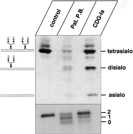
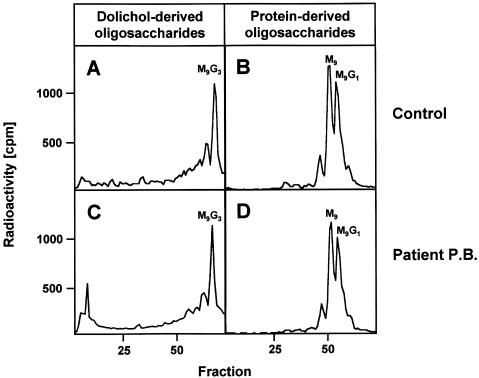
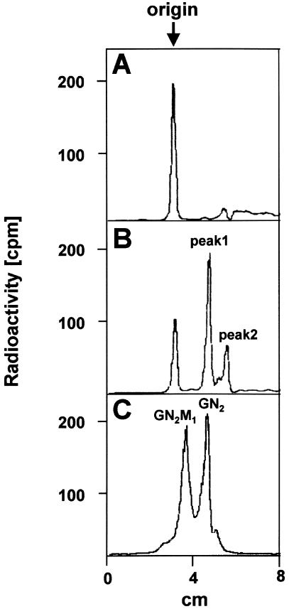
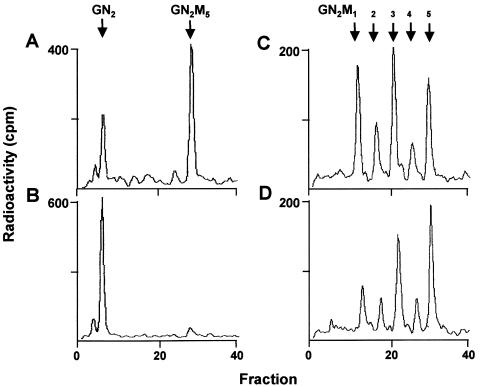
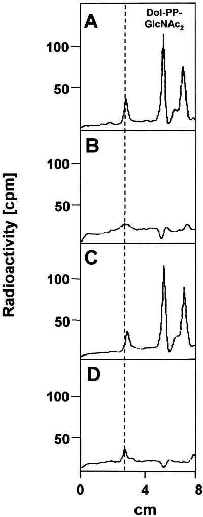
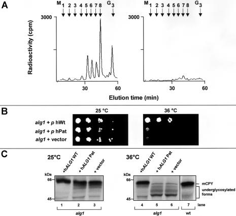
The molecular nature of a severe multisystemic disorder with a recurrent nonimmune hydrops fetalis was identified as deficiency of GDP-Man:GlcNAc(2)-PP-dolichol mannosyltransferase, the human orthologue of the yeast ALG1 gene (MIM 605907). The disease belongs to the group of congenital disorders of glycosylation (CDG) and is designated as subtype CDG-Ik. In patient-derived serum, the total amount of the glycoprotein transferrin was reduced. Moreover, a partial loss of N-glycan chains was observed, a characteristic feature of CDG type I forms. Metabolic labeling with [6-(3)H]glucosamine revealed an accumulation of GlcNAc(2)-PP-dolichol and GlcNAc(1)-PP-dolichol in skin fibroblasts of the patient. Incubation of fibroblast extracts with [(14)C]GlcNAc(2)-PP-dolichol and GDP-mannose indicated a severely reduced activity of the beta 1,4-mannosyltransferase, elongating GlcNAc(2)-PP-dolichol to Man(1)GlcNAc(2)-PP-dolichol at the cytosolic side of the endoplasmic reticulum. Genetic analysis of the patient's hALG1 gene identified a homozygous mutation leading to the exchange of a serine residue to leucine at position 258 in the hALG1 protein. The disease-causing nature of the hALG1 mutation for the glycosylation defect was verified by a retroviral complementation approach in patient-derived primary fibroblasts and was confirmed by the expression of wild-type and mutant hALG1 in the Saccharomyces cerevisiae alg1-1 strain.


Figures

Figure  1
Figure 1
IEF pattern and SDS-PAGE of serum transferrin. Sera from a control, patient P.B., and a patient with CDG-Ia were analyzed by IEF (upper panel) and SDS-PAGE, followed by western blotting (lower panel) and immunodetection of transferrin. “Tetrasialo,” “disialo,” and “asialo” on the upper panel indicate transferrin forms with four, two, or no sialic acid residues. The numerals “2,” “1,” and “0” in the lower panel indicate transferrin forms with two, one, or zero oligosaccharide chains.
Figure  2
Figure 2
Analysis of protein- and dolichol-derived oligosaccharides in CDG-Ik. Fibroblasts of a control (B) and the patient (D) were metabolically labeled with [2-3H]mannose for 30 min, [2-3H]glycans were released from equal amounts of the glycoprotein fraction by PNGase F digestion and were size fractionated by HPLC. M9 and M9G1 refer to the positions of GlcNAc2Man9 and GlcNAc2Man9Glc1 standards, respectively. Control- (A) and patient-derived (C) fibroblasts (in equal amounts) were metabolically labeled with [2-3H]mannose for 30 min. The [2-3H]oligosaccharides were released from the dolichol-PP moiety by mild acid hydrolysis and were size fractionated by HPLC. M9G3 refers to the position of a GlcNAc2Man9Glc3 standard.
Figure  3
Figure 3
TLC analysis of [6-3H]glucosamine-labeled short dolichol-linked oligosaccharides. Fibroblasts from a control (A) and from patient P.B. (B) were metabolically labeled for 60 min with [6-3H]glucosamine. After extraction of the short lipid-linked oligosaccharide fraction with chloroform:methanol (3:2), further analysis was performed by TLC on silica gel 60 plates with chloroform:methanol:water (65:25:4) as solvent. The position of the origin and the positions of a [14C]GlcNAc2-PP-dolichol standard and a Man1[14C]GlcNAc2-PP-dolichol standard are indicated as GN2 and GN2M1, respectively (C).
Figure  4
Figure 4
In vitro determination of hALG1 and hALG2 activity. Microsomal extracts from fibroblasts of a control (A, C) and the patient (B, D) were incubated for 10 min with either [14C]GlcNAc2-PP-dolichol (A, B) or with Man1[14C]GlcNAc2-PP-dolichol (C, D), respectively, in the presence of GDP-mannose. Dolichol-linked oligosaccharides were extracted from the incubation mixture and were treated by mild acid hydrolysis, and the released oligosaccharides were separated by HPLC. The positions of a GlcNAc2-standard (GN2) and Man1–5GlcNAc2 (GN2M1–5)-standards are marked by arrows.
Figure  5
Figure 5
Retroviral transduction of patient fibroblasts with wild-type hALG1 cDNA leads to complementation of the hALG1 deficiency. Dolichol-PP-[3H]GlcNAc2 and dolichol-PP-[3H]GlcNAc1 were extracted with chloroform:methanol (3:2) from [6-3H]glucosamine-labeled control fibroblasts expressing the retroviral vector alone (D) and from patient fibroblasts, which were either transduced with the retroviral vector alone (A), the wild-type cDNA (B), or the C773T hALG1 cDNA (C). Further analysis was performed by TLC. The elution position of a [14C]GlcNAc2-PP-dolichol standard and the origin (dotted line) are indicated.
Figure  6
Figure 6
Defects in dolichol-linked oligosaccharide biosynthesis (A), growth (B), and CPY glycosylation (C) in the alg1-1 yeast mutant are complemented by expression of wild-type hALG1. A, Biosynthesis of dolichol-linked oligosaccharides was investigated in an alg1-1 strain transformed with the wild-type hALG1 cDNA (left panel) or the hALG1 cDNA encoding the C773T mutation (right panel). Yeast cells were metabolically labeled with [2-3H]mannose for 30 min, [2-3H] oligosaccharides were released from the dolichol moiety by mild acid hydrolysis and further analyzed by HPLC. M1–M8 and G3 refer to Man1–8GlcNAc2 and Glc3Man9GlcNAc2 standards, respectively. B, Growth of yeast alg1-1 cells either transformed with wild-type hALG1 cDNA, the C773T hALG1 or the expression vector was investigated under permissive (25°C, left panel) and nonpermissive temperature (36°C, right panel). C, The glycosylation status of CPY is shown at the permissive temperature (25°C, left panel) and at the nonpermissive temperature (36°C, right panel). Yeast cells were metabolically labeled with 35S-methionine for 30 min, and CPY was immunoprecipitated and analyzed by SDS-PAGE. The position of the mature form of CPY in wild-type yeast cells (mCPY) and in the complemented alg1-1 cells are indicated on the right. Molecular weight standards are indicated on the left.

References

Electronic-Database Information

    1. National Center for Biotechnology Information (NCBI) Entrez Database, http://www.ncbi.nlm.nih.gov/Entrez/ (for hALG1 cDNA [accession number BAA90748])
    1. Online Mendelian Inheritance in Man (OMIM), http://www.ncbi.nlm.nih.gov/Omim/ (for ALG1 and CDG-Ii)

References

    1. Aebi M, Helenius A, Schenk B, Barone R, Fiumara A, Berger EG, Hennet T, et al (1999) Carbohydrate-deficient glycoprotein syndromes become congenital disorders of glycosylation: an updated nomenclature for CDG. First International Workshop on CDGS. Glycoconj J 16:669–67110.1023/A:1017249723165 - DOI - PubMed
    1. Gietz RD, Schiestl RH (1991) Applications of high efficiency lithium acetate transformation of intact yeast cells using single stranded nucleic acids as carrier. Yeast 7:253–263 - PubMed
    1. Helenius A, Aebi A (2001) Intracellular functions of N-linked glycans. Science 291:2364–236910.1126/science.291.5512.2364 - DOI - PubMed
    1. Huffaker T, Robbins P (1982) Temperature-sensitive yeast mutants deficient in asparagine-linked glycosylation. J Biol Chem 257:3203–3210 - PubMed
    1. Jaeken J (2003) Congenital disorders of glycosylation (CDG): it’s all in it! J Inherit Metab Dis 26:99–11810.1023/A:1024431131208 - DOI - PubMed

Publication types

MeSH terms

Substances