. 2004 Mar;134(3):1206-16.
doi: 10.1104/pp.103.035832. Epub 2004 Feb 19.

Plastid lysophosphatidyl acyltransferase is essential for embryo development in Arabidopsis

Affiliations

Plastid lysophosphatidyl acyltransferase is essential for embryo development in Arabidopsis

Hyun Uk Kim et al. Plant Physiol. 2004 Mar.

Abstract

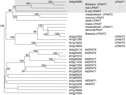
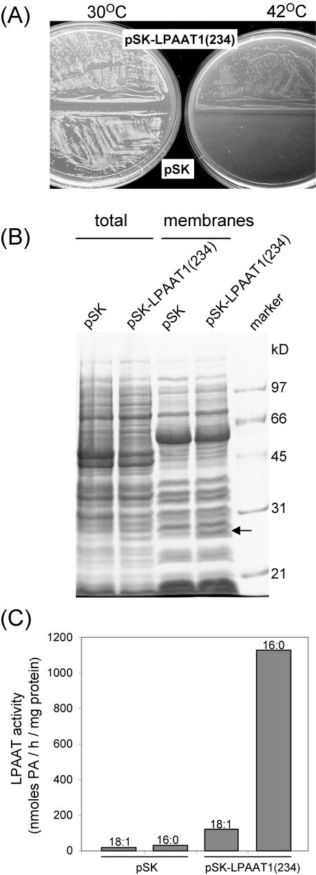
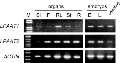
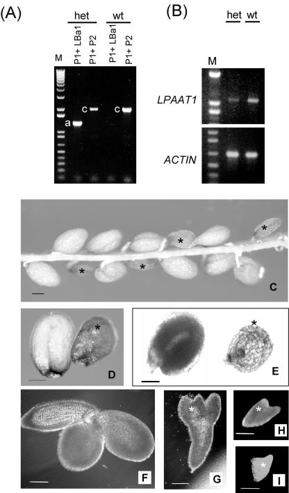
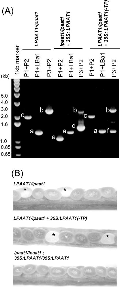
Lysophosphatidyl acyltransferase (LPAAT) is a pivotal enzyme controlling the metabolic flow of lysophosphatidic acid into different phosphatidic acids in diverse tissues. A search of the Arabidopsis genome database revealed five genes that could encode LPAAT-like proteins. We identified one of them, LPAAT1, to be the lone gene that encodes the plastid LPAAT. LPAAT1 could functionally complement a bacterial mutant that has defective LPAAT. Bacteria transformed with LPAAT1 produced LPAAT that had in vitro enzyme activity much higher on 16:0-coenzyme A than on 18:1-coenzyme A in the presence of 18:1-lysophosphatidic acid. LPAAT1 transcript was present in diverse organs, with the highest level in green leaves. A mutant having a T-DNA inserted into LPAAT1 was identified. The heterozygous mutant has no overt phenotype, and its leaf acyl composition is similar to that of the wild type. Selfing of a heterozygous mutant produced normal-sized and shrunken seeds in the Mendelian ratio of 3:1, and the shrunken seeds could not germinate. The shrunken seeds apparently were homozygous of the T-DNA-inserted LPAAT1, and development of the embryo within them was arrested at the heart-torpedo stage. This embryo lethality could be rescued by transformation of the heterozygous mutant with a 35S:LPAAT1 construct. The current findings of embryo death in the homozygous knockout mutant of the plastid LPAAT contrasts with earlier findings of a normal phenotype in the homozygous mutant deficient of the plastid glycerol-3-phosphate acyltransferase; both mutations block the synthesis of plastid phosphatidic acid. Reasons for the discrepancy between the contrasting phenotypes of the two mutants are discussed.


Figures

Figure 1.
Figure 1.
A phylogenetic tree of Arabidopsis genes that encode proteins related to LPAAT constructed on the basis of their predicted amino acid sequences. It was inferred from the alignment using the neighbor-joining method with 1,000 bootstrap replicates. Only bootstrap values of over 50% are shown. Genes encoding these proteins were obtained after a BLAST search of the databases of The Arabidopsis Information Resource and National Center for Biotechnology Information with the use of the amino acid sequences of a maize cytoplasmic LPAAT (Z29518) and a B. napus plastid LPAAT1 (AF111161) as queries (for details, see “Results”). All of the preceding and following numbers of genes/proteins are from GenBank. Eleven putative LPAATs cited in a Web site (Beisson et al., 2003) and studied ATs (GPAT and DGAT1) of the Kennedy pathway are included. All reported LPAATs of other plant species (rice, AC068923; meadowfoam LPAAT2, S60477; coconut, U29657; meadowfoam LPAAT1, S60478; almond, AF213937; and B. napus LPAAT2, Z95637) and Escherichia coli (from plsC, M63491) and yeast (Saccharomyces cerevisiae) LPAAT (from slc1, L13282) are incorporated. The Arabidopsis genes are shown by their locus numbers, and the first five genes are also shown by their simplified protein names, as are those from other plants and microbes. Arabidopsis LPAAT1, B. napus plastid LPAAT1, a rice LPAAT (presumably in the plastids), and E. coli LPAAT are shaded.
Figure 2.
Figure 2.
A comparison of the structures of the Arabidopsis LPAAT1 and putative cytoplasmic LPAAT(2–5) and those of the B. napus plastid LPAAT1 and cytoplasmic LPAAT2. The Arabidopsis (LPAAT1) and B. napus (Bn LPAAT1) plastid enzymes are similar in the locations of the putative plastid transit peptide (TP), the two trans-membrane segments (wide vertical bars), and the successive conserved motifs NHX4D and EGT (narrow vertical bars). LPAAT2 to 5 and the B. napus cytoplasmic LPAAT (Bn LPAAT2) share similar locations of the trans-membrane segments and the successive conserved motifs and an absence of a putative plastid transit peptide. The beginning residue numbers of the various parameters along the sequences are indicated.
Figure 3.
Figure 3.
Structure of Arabidopsis LPAAT1 (At4g30580) encoding plastid LPAAT1, lpaat1, and the derived constructs used in the current studies. A 2-kb segment of the gene possessing seven exons, which encode the putative N-terminal plastid targeting signal (white boxes) and the mature protein (shaded boxes), is shown. The mutated gene, lpaat1, is interrupted by a T-DNA (indicated with an inserted triangle). The horizontal arrow along the ORF represents the starting point of the LPAAT1(234) segment, which was used to transform E. coli for testing functional complementation and enzyme activity. The structures of 35S:LPAAT1 and 35S:LPAAT1(-TP), which were used to transform Arabidopsis for testing functional complementation, are shown as horizontal boxes at the lower portion. The horizontal lines labeled a to f denote the predicted PCR fragments produced from the various primers. The primers correspond to the 5′ (P1) and 3′ (P2) termini of LPAAT1 or the left (LBa1) and right (P3) borders of the T-DNA. The 35S (heavily shaded box) also contains the P3 sequence.
Figure 4.
Figure 4.
Expression of At LPAAT1 in E. coli strain JC201 temperature-sensitive mutant defective of LPAAT. A, Bacteria transformed with pSK or pSK-LPAAT1(234) were grown at 30°C or 42°C for 18 h and photographed. B, Total extracts and the membrane fractions of the two types of transformed bacteria grown at 30°C were analyzed for their protein constituents by SDS-PAGE. A protein of 28 kD (arrowed) was present in the total extract and the membrane fraction of bacteria transformed with pSK-LPAAT1(234) but absent in those of bacteria transformed with pSK. Mr markers are on the right lane. C, LPAAT activities in the membrane fractions from the two types of transformed bacteria assayed with the use of equal amounts of proteins (30 μg), LPA-18:1, and either 18:1- or 16:0-CoA are shown.
Figure 5.
Figure 5.
RT-PCR analysis of the transcript from various organs with primers specific for LPAAT1 or LPAAT2. Approximately equal amounts of transcript of an Arabidopsis actin gene (ACTIN) were present in the various samples. Organs included maturing siliques (Si), maturing flowers (F), rosette leaves (RL), stems (St), and roots (R), as well as early (E) and late (L) maturing embryos and seedlings. Left lane, Markers of DNA length.
Figure 6.
Figure 6.
Characterization of the LPAAT1/lpaat1 heterozygous Arabidopsis mutant. A, PCR products of LPAAT1 and lpaat1 with the use of leaf DNA of wild type (wt) and a heterozygous LPAAT1/lpaat1 mutant (het). The primers representing the 5′ (P1) and 3′ (P2) termini of LPAAT1 (producing PCR fragment c) or P1 and the left border of the T-DNA (LBa1; producing PCR fragment a) are shown in Figure 3. Left lane, DNA size markers. B, RT-PCR products of LPAAT1 with the use of equal amounts of leaf RNA and the primers P1 and P2. The reaction detected LPAAT1 but not lpaat1 because of the exceedingly long, inserted T-DNA (approximately 5 kb). Approximately equal amounts of transcript of an Arabidopsis ACTIN gene were present in the two samples. DNA size markers are in the left lanes. C to I, Light microscopic images of the seeds and embryos produced by a LPAAT1/lpaat1 heterozygous mutant. C, Almost mature (approximately 15 d after flowering [DAF]) silique with its coat removed to reveal normal-sized and shrunken (asterisk) seeds. D, Enlarged view of normal-sized and a shrunken seeds taken from an almost mature (approximately 15 DAF) silique observed under a dissecting microscope. E, Same as D except under a transmission microscope. Both types of microscopy revealed that the normal-sized seed was completely filled with an upturned-U embryo, whereas the shrunken seed contained a very small embryo. F, Spread-out embryo from a normalsized seed of approximately 12 DAF. G to I, Spread-out embryos of different sizes from individual shrunken seeds of approximately 12 DAF. Bar in all images = 100 μm.
Figure 7.
Figure 7.
Functional complementation of Arabidopsis lpaat1 mutants with LPAAT1. A, PCR products from leaf DNA of heterozygous (LPAAT1/lpaat1) or homozygous lpaat1/lpaat1) mutants as offspring of heterozygous plants that have or have not been transformed with 35S:LPAAT1 or 35S:LPAAT1(-TP). The structures of these constructs and the expected PCR fragments a to f derived from the various primers are shown in Figure 3. Left lane, DNA size markers. B, Light microscopic images of the maturing (approximately 12 DAF) seeds in siliques in plants of the indicated genotypes. Stars indicate white, transparent maturing seeds, which would become shrunken upon silique maturation.

Similar articles

Cited by

References

    1. Beisson F, Koo AJK, Ruuska S, Schwender J, Pollard M, Thelen J, Paddock T, Salas J, Savage L, Milcamps A et al. (2003) Arabidopsis thaliana genes involved in acyl lipid metabolism: a 2003 census of the candidates, a study of the distribution of ESTs in organs and a Web-based database. Plant Physiol 132: 681-697 - PMC - PubMed
    1. Bourgis F, Kader JC, Barret P, Renard M, Robinson D, Robinson C, Delseny M, Roscoe TJ (1999) A plastidal lysophosphatidic acid acyltransferase from oilseed rape. Plant Physiol 120: 913-921 - PMC - PubMed
    1. Bouvier-Nave P, Benveniste P, Oelkers P, Sturley SL, Schaller H (2000) Expression in yeast and tobacco of plant cDNAs encoding acyl CoA: diacylglycerol acyltransferase. Eur J Biochem 267: 85-96 - PubMed
    1. Brown AP, Brough CL, Kroon JTM, Slabas AR (1995) Identification of a cDNA that encodes a 1-acyl-sn-glycerol-3-phosphate acyltransferase from Limnanthes douglasii. Plant Mol Biol 29: 267-278 - PubMed
    1. Brown AP, Coleman J, Tommey AM, Watson MD, Slabas AR (1994) Isolation and characterization of a maize cDNA that complements a 1-acyl-sn-glycerol-3-phosphate acyltaransferase mutant of Escherichia coli and encodes a protein which similarities to other acyltransferases. Plant Mol Biol 26: 211-223 - PubMed

Publication types

MeSH terms