. 2004 Mar 2;101(9):3005-10.
doi: 10.1073/pnas.0308369101. Epub 2004 Feb 19.

Intact type 1 immunity and immune-associated coagulative responses in mice lacking IFN gamma-inducible fibrinogen-like protein 2

Affiliations

Intact type 1 immunity and immune-associated coagulative responses in mice lacking IFN gamma-inducible fibrinogen-like protein 2

Wayne W Hancock et al. Proc Natl Acad Sci U S A. .

Abstract

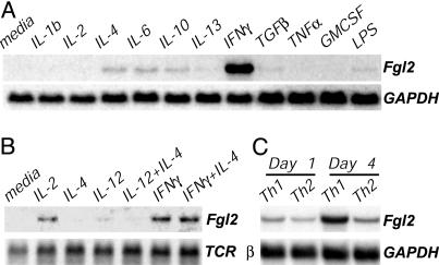
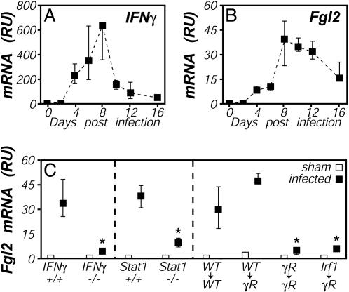
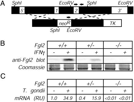
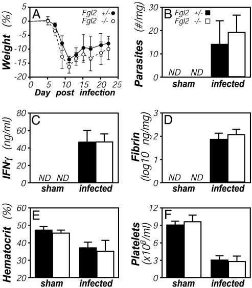
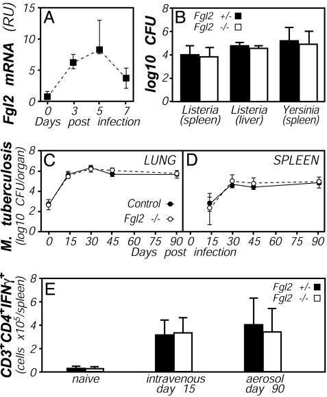
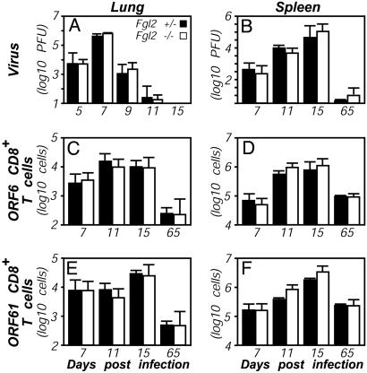
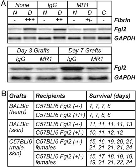
Fibrinogen-like protein 2 (Fgl2, fibroleukin) is a leukocyte product that exhibits significant homology to secreted proteins of diverse function, including growth factors, lectins, and components of extracellular matrix. Prior studies found that Fgl2 is IFN gamma-inducible, possesses direct coagulant activity, and inhibits T cell proliferation and dendritic cell maturation in vitro. Here, we demonstrate that Fgl2 expression is up-regulated during type 1 immunity in vivo and establish that such up-regulation is IFN gamma-, signal transducer and activation of transcription protein 1-, and IFN response factor 1-dependent. To investigate functional roles for Fgl2 during type 1 immunity, we generated Fgl2-deficient mice. Those animals are born at predicted Mendelian frequencies, appear overtly healthy, and contain normal numbers and frequencies of lymphoid cells. Although Fgl2 is IFN gamma-inducible and putatively regulates T cell activation/proliferation, we demonstrate that Fgl2-deficient and control mice exhibit similar degrees of T cell expansion, immunopathology, and/or pathogen burdens during protozoan (Toxoplasma gondii), bacterial (Yersinia enterocolitica, Listeria monocytogenes, and Mycobacterium tuberculosis), and viral (murine gamma-herpesvirus-68 and Sendai) infections. Fgl2-deficient mice also reject allografts with similar kinetics as control mice. Moreover, despite prior reports that Fgl2 functions as a procoagulant enzyme, we demonstrate that Fgl2-deficient and control mice produce similar levels of fibrin, a product of the coagulation cascade, during T. gondii infection and allograft rejection. Together, our findings suggest that Fgl2, although highly conserved and IFN gamma-inducible, is not a critical mediator of either type 1 immunity or immune-associated coagulant activity.


Figures

Fig. 1.
Fig. 1.
IFNγ-inducible and T helper (Th)1-biased expression of Fgl2 in vitro. Northern blots are shown. (A) RAW264.7 mouse macrophages stimulated for 6 h with the indicated cytokines (5 ng/ml) or lipopolysaccharide (10 μg/ml). (B) C57BL/6 lymph node cells cultured for 24 h in the presence of the indicated cytokines. (C) C57BL/6 lymph node cells stimulated with plate-bound anti-CD3 in the presence of IL-12 and anti-IL-4 (Th1 conditions), or IL-4 and anti-IFNγ (Th2 conditions).
Fig. 2.
Fig. 2.
IFNγ-, Stat1-, and Irf-dependent up-regulation of Fgl2 expression during T. gondii infection. (A and B) Mice were perorally infected with 10 ME49 T. gondii cysts, and tissues were harvested on the indicated days. Relative levels of mRNA encoding IFNγ (A) and Fgl2 (B) were determined by real-time PCR. (C) Mice were infected with T. gondii (filled symbols) or were sham-infected (open symbols). Tissues were harvested on day 8 after infection, and relative levels of Fgl2 mRNA were determined by real-time PCR. (Right) Shown is an experiment using bone marrow chimeras in which WT, IFNγR-deficient, or Irf1-deficient bone marrow was used to reconstitute lethally irradiated WT or IFNγR-deficient hosts. Throughout, data depict the median and range of four to five mice per time point. *, P < 0.05.
Fig. 3.
Fig. 3.
Production of Fgl2-deficient mice. (A Upper) Schematic of the Fgl2 gene, indicating (i) locations of the two Fgl2 exons, (ii) restriction enzyme sites and probes used for confirming successful disruption of the fgl2 gene by Southern blotting, and (iii) locations of PCR primers (arrows) used to routinely screen for the targeted mutation. (Lower) Schematic of the targeting construct, indicating regions flanking the fgl2 exons that were cloned into the targeting vector and the relative placement of selectable elements. neoR, neomycin resistance gene; TK, thymidine kinase gene. (B) Impaired secretion of Fgl2 protein by Fgl2-deficient cells. Thioglycollate-elicited peritoneal macrophages were isolated from littermate mice of the indicated fgl2 genotype. After 24 h of culture in the presence or absence of IFNγ (20 ng/ml), cell-free supernatants were assayed for Fgl2 expression by Western blotting with Fgl2-specific mAb. Coomassie blue staining of a gel run in parallel documented equivalent sample loading in all lanes. (C) Impaired production of Fgl2 mRNA by T. gondii-infected Fgl2-deficient mice. As in Fig. 2, mice were infected with T. gondii, liver tissue was harvested on day 8 after infection, and levels of Fgl2 mRNA were determined by real-time PCR. Data depict the average of four to five mice per group after normalization to uninfected WT mice.
Fig. 4.
Fig. 4.
T. gondii infection of Fgl2-deficient mice. Fgl2-deficient (open bars) and littermate control Fgl2-heterozygous (filled bars) mice were infected with T. gondii, as in Fig. 2. (A) All mice survived the acute phase of the infection, and there were no significant differences in weight loss/recovery (n = 5, P > 0.05 at all time points). At day 8 after infection, cohorts of five mice were assayed for hepatic parasite burdens by real-time PCR (B), levels of IFNγ protein in plasma by ELISA (C), and levels of hepatic fibrin by quantitative Western blotting (D). In addition, hematocrits (E) and platelet numbers (F) were quantified by Coulter Counter. None of the measured parameters differed significantly between Fgl2-deficient and heterozygous control mice.
Fig. 5.
Fig. 5.
Bacterial responses in Fgl2-deficient mice. (A) Up-regulation of Fgl2 expression during bacterial infection. WT C57BL/6 mice were i.p. infected with 1 × 10⁵ L. monocytogenes, and levels of Fgl2 mRNA levels were quantified by real-time PCR. (B) Similar bacterial burdens in Fgl2-deficient (open bars) and control heterozygous mice (filled bars) after bacterial infection. Colony-forming units in the indicated organs were determined at day 5 after i.p. infection with 1 × 10⁵ L. monocytogenes or i.v. infection with 5 × 10³ Y. enterocolitica. In each case, data depict average and standard deviation of five mice per group, and no statistically significant differences were observed. (C-E) Similar bacterial burdens and T cell responses in Fgl2-deficient (open symbols) and control mice (filled symbols) after mycobacterial infection. CFUs in lung (C) or spleen (D) were determined on the indicated days after aerosol infection with M. tuberculosis. (E) Numbers of splenic CD3+CD4+ cells that could produce IFNγ on in vitro reactivation were quantified by flow cytometry 15 days after i.v. infection or 90 days after aerosol infection with M. tuberculosis. For C-E, data depict the average and standard deviation of four mice per group, except that only two Fgl2-deficient mice were analyzed at day 45.
Fig. 6.
Fig. 6.
Antiviral responses in Fgl2-deficient mice. (A and B) Similar viral titers in Fgl2-deficient (open bars) and heterozygous control (filled bars) mice. Mice were intranasally infected with 400 plaque-forming units (pfus) MHV-68. Viral titers in the lung (A) and spleen (B) were determined at the indicated times after infection. The detection limit of the plaque assay was 1 pfu per 1.2 × 10⁷ spleen cells. Data shown are representative of two independent experiments. (C-F) Similar numbers of virus-specific T cells in Fgl2-deficient (open bars) and heterozygous control (filled bars) mice. MHV-68-specific CD8+ T cells in lung airways (C and E) and the spleen (D and F) were quantified by flow cytometry after staining with MHC class I tetramer ORF6487-495/Db (C and D) or ORF61524-531/Kb (E and F). Data depict the average and standard deviation of three mice per time point.
Fig. 7.
Fig. 7.
Expression and function of Fgl2 during graft rejection. (A) Expression of Fgl2 mRNA correlates with fibrin deposition and allograft rejection. (Upper) Heterotopic BALB/c cardiac allografts were transplanted to C57BL/6 mice. Northern blots were prepared from day 3 donor (D) BALB/c allograft hearts, native (N) C57BL/6 hearts from the allograft recipients, and control (C) nontransplanted BALB/c hearts. Blots were successively hybridized with the indicated probes. Graft recipients received no treatment (None), donor spleen cell transfusion plus IgG, or donor spleen cell transfusion plus anti-CD40 ligand mAb MR1, as indicated. Fibrin was measured semiquantitatively by immunohistochemistry. (Lower) A second Northern blot was prepared from an independent series of day 3 and day 7 graft samples. (B) Similar kinetics of graft rejection in Fgl2-deficient and heterozygous control mice. As indicated, cardiac or skin grafts were transplanted to Fgl2-deficient or control mice, and times to rejection were recorded.

References

    1. Ruegg, C. & Pytela, R. (1995) Gene 160, 257-262. - PubMed
    1. Marazzi, S., Blum, S., Hartmann, R., Gundersen, D., Schreyer, M., Argraves, S., von Fliedner, V., Pytela, R. & Ruegg, C. (1998) J. Immunol. 161, 138-147. - PubMed
    1. Koyama, T., Hall, L. R., Haser, W. G., Tonegawa, S. & Saito, H. (1987) Proc. Natl. Acad. Sci. USA 84, 1609-1613. - PMC - PubMed
    1. Lafuse, W. P., Castle, L., Brown, D. & Zwilling, B. S. (1995) Cell. Immunol. 163, 187-190. - PubMed
    1. Parr, R. L., Fung, L., Reneker, J., Myers-Mason, N., Leibowitz, J. L. & Levy, G. (1995) J. Virol. 69, 5033-5038. - PMC - PubMed

Publication types