. 2004 Mar 2;101(9):2894-9.
doi: 10.1073/pnas.0305252101. Epub 2004 Feb 19.

Disruption of TRPM6/TRPM7 complex formation by a mutation in the TRPM6 gene causes hypomagnesemia with secondary hypocalcemia

Affiliations

Disruption of TRPM6/TRPM7 complex formation by a mutation in the TRPM6 gene causes hypomagnesemia with secondary hypocalcemia

Vladimir Chubanov et al. Proc Natl Acad Sci U S A. .

Abstract

Impaired magnesium reabsorption in patients with TRPM6 gene mutations stresses an important role of TRPM6 (melastatin-related TRP cation channel) in epithelial magnesium transport. While attempting to isolate full-length TRPM6, we found that the human TRPM6 gene encodes multiple mRNA isoforms. Full-length TRPM6 variants failed to form functional channel complexes because they were retained intracellularly on heterologous expression in HEK 293 cells and Xenopus oocytes. However, TRPM6 specifically interacted with its closest homolog, the Mg(2+)-permeable cation channel TRPM7, resulting in the assembly of functional TRPM6/TRPM7 complexes at the cell surface. The naturally occurring S141L TRPM6 missense mutation abrogated the oligomeric assembly of TRPM6, thus providing a cell biological explanation for the human disease. Together, our data suggest an important contribution of TRPM6/TRPM7 heterooligomerization for the biological role of TRPM6 in epithelial magnesium absorption.


Figures

Fig. 1.
Fig. 1.
Human TRPM6 gene structure and expression pattern of TRPM6 and TRPM7. (A) Intron–exon structure of the human TRPM6 gene according to the primary sequence analysis of cDNA variants cloned. Exons encoding putative transmembrane domains (S1–6) and the predicted kinase domain are highlighted by a gray background. Exon 36B, spliced between exon 36 and 37, was detected only in mRNA from human testis. (B) Diagnostic RT-PCR for TRPM6a, TRPM6b, and TRPM7 mRNA expression in HEK 293, H69, and H510 small cell lung carcinoma cells as well as in human bronchial epithelial cells (BEAS-2B), human bronchial smooth muscle cells (BSMC), human testis, and human kidney. (C) Diagnostic TRPM7 RT-PCR on microdissected rat nephron segments, including glomeruli (glom), proximal convoluted tubule (PCT), descending thin limb (dTL), ascending TL (aTL), medullary thick ascending limb (mTAL), cortical TAL (cTAL), distal convoluted tubule DCT, connecting tubule/cortical collecting duct (CT/CCD), outer medullary collecting duct (OMCD), and inner medullary collecting duct (IMCD).
Fig. 2.
Fig. 2.
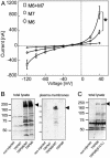
Heterologous expression of TRPM6/7 in Xenopus oocytes. (A) Two-electrode voltage clamp analysis of oocytes injected with 10 ng of cRNA encoding TRPM6 (M6), TRPM7 (M7), or 10 ng of TRPM6 together with 10 ng of TRPM7 cRNA (M6+M7). IV relationships were determined on at least seven oocytes per IV curve. *, P < 0.05. (B) Western blot analysis of TRPM6 expression in total lysates (Left) and in plasma membranes (Right) of Xenopus oocytes, using a TRPM6-specific antiserum. (C) Western blot analysis of TRPM6 and TRPM6(S141L) expression in total lysates of Xenopus oocytes. The positions of TRPM6-specific bands are indicated by arrowheads.
Fig. 3.
Fig. 3.
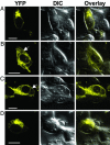
Subcellular localization of transiently expressed TRPM6 and TRPM7 in living HEK 293 cells. TRPM6 and TRPM7 were C-terminally fused to YFP and transiently expressed in HEK 293 cells. TRPM6-YFP (A) and TRPM7-YFP (B) were expressed alone; TRPM6-YFP (C) and TRPM6(S141L)-YFP (D) were coexpressed with untagged TRPM7 at a ratio of 1:3 (YFP-tagged/untagged cDNAs). Images of YFP fluorescence (YFP), the corresponding differential interference contrast images (DIC), and overlays of both are depicted. (Scale bars, 10 μm.) The arrow indicates the plasma membrane. Typical examples of three independent transfections are shown.
Fig. 4.
Fig. 4.
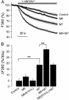
TRPM6- and TRPM7-mediated Mn2+ entry in HEK 293 cells. HEK 293 cells were transiently cotransfected with plasmids (50 ng) encoding GFP and a mixture of plasmid DNAs as indicated: Control, 2 μg of empty pcDNA3.1 vector; M6, 1 μg of TRPM6 plus 1 μg of pcDNA3.1; M7, 1 μg of TRPM7 plus 1μg of pcDNA3.1; M6+M7, 1 μg of TRPM6 plus 1 μg of TRPM7; M6(S141L)+M7, 1 μg of TRPM6(S141L) plus 1 μg of TRPM7. Bold black lines indicate calculated mean values of individual traces recorded from single cells, and gray lines depict the SEM obtained for each time point. Addition of 1 mM Mn2+ in HBS is indicated by a gray bar. Typical examples from four independent transfections are shown. (B) Rate of Mn2+ entry (percentage decrease of fluorescence at 360 nm over 30 sec) is presented as means and SEM calculated from four independent transfections. **, P < 0.01.
Fig. 5.
Fig. 5.
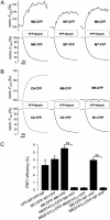
TRPM6/7 assembly as assessed by FRET. (A) TRPM6 and TRPM7 C-terminally fused to CFP or YFP were coexpressed in HEK 293 cells. Static FRET signals were recorded between TRPM6-CFP/YFP (Left), TRPM7-CFP/YFP (Center), and TRPM6-CFP/TRPM7-YFP (Right). (B) Static FRET between TRPC6-YFP/TRPC6-CFP subunits was used as a positive control (Left). Other tested FRET combinations were TRPM6-CFP/TRPC6-YFP (Center) and TRPM6-CFP/TRPM1-YFP (Right). (C) Mean values and SEM of FRET efficiency were calculated from four independent transfections. **, P < 0.01.

Similar articles

Cited by

References

    1. Hardie, R. C. (2001) J. Exp. Biol. 204, 3403-3409. - PubMed
    1. Clapham, D. E., Montell, C., Schultz, G. & Julius, D. (2003) Pharmacol. Rev. 55, 591-596. - PubMed
    1. Clapham, D. E. (2003) Nature 426, 517-524. - PubMed
    1. Voets, T. & Nilius, B. (2003) J. Membr. Biol. 192, 1-8. - PubMed
    1. Hofmann, T., Schaefer, M., Schultz, G. & Gudermann, T. (2002) Proc. Natl. Acad. Sci. USA 99, 7461-7466. - PMC - PubMed

Publication types

MeSH terms