. 2004 Mar 10;23(5):1008-19.
doi: 10.1038/sj.emboj.7600102. Epub 2004 Feb 19.

Domain movements of elongation factor eEF2 and the eukaryotic 80S ribosome facilitate tRNA translocation

Affiliations

Domain movements of elongation factor eEF2 and the eukaryotic 80S ribosome facilitate tRNA translocation

Christian M T Spahn et al. EMBO J. .

Abstract

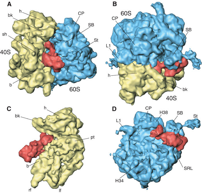
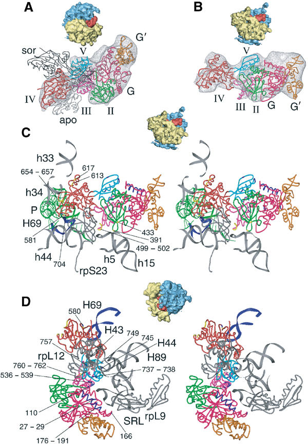
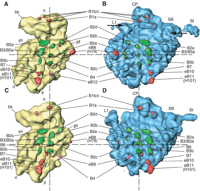
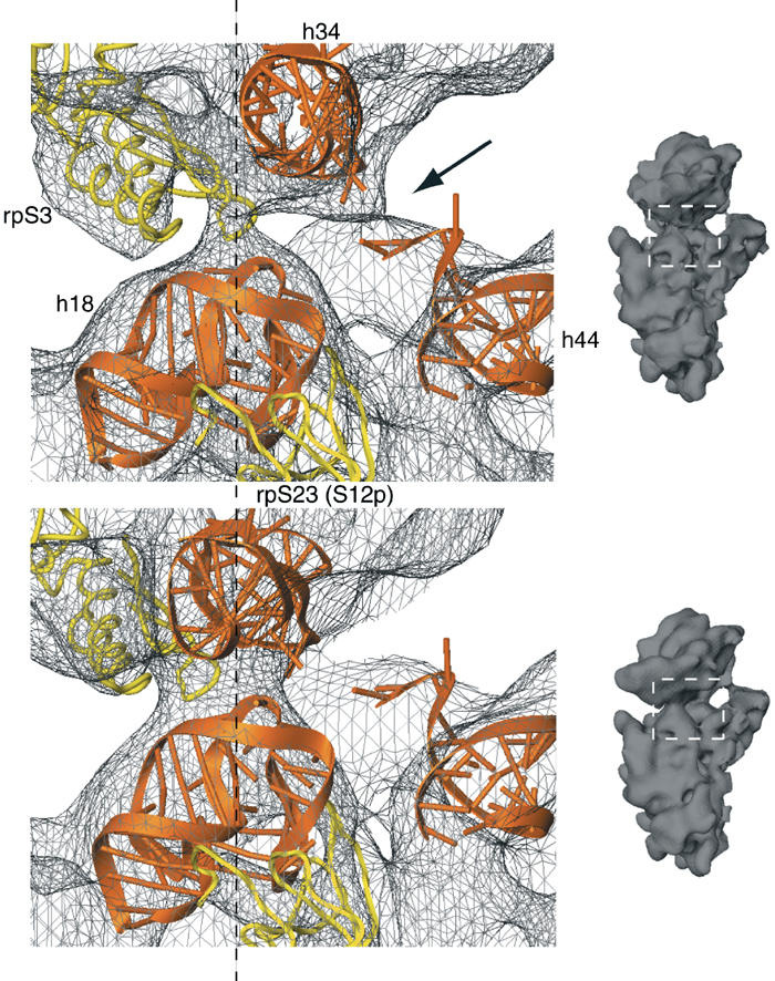
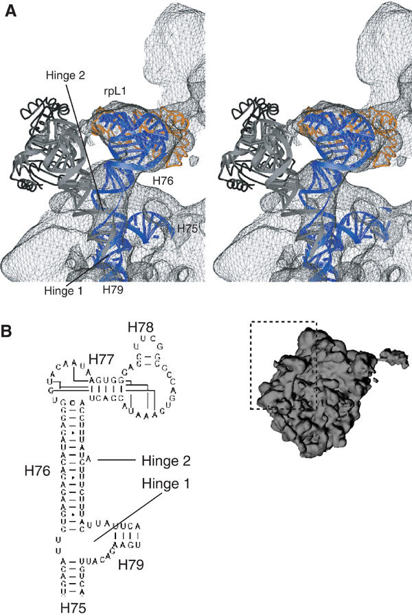
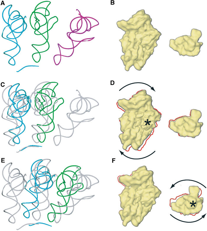
An 11.7-A-resolution cryo-EM map of the yeast 80S.eEF2 complex in the presence of the antibiotic sordarin was interpreted in molecular terms, revealing large conformational changes within eEF2 and the 80S ribosome, including a rearrangement of the functionally important ribosomal intersubunit bridges. Sordarin positions domain III of eEF2 so that it can interact with the sarcin-ricin loop of 25S rRNA and protein rpS23 (S12p). This particular conformation explains the inhibitory action of sordarin and suggests that eEF2 is stalled on the 80S ribosome in a conformation that has similarities with the GTPase activation state. A ratchet-like subunit rearrangement (RSR) occurs in the 80S.eEF2.sordarin complex that, in contrast to Escherichia coli 70S ribosomes, is also present in vacant 80S ribosomes. A model is suggested, according to which the RSR is part of a mechanism for moving the tRNAs during the translocation reaction.


Figures

Figure 1
Figure 1
A 11.7-Å-resolution cryo-EM map of the yeast 80S·eEF2·sordarin complex. The cryo-EM map is shown (A) from the side; (B) from the top ; (C) from the 60S side, with 60S removed; and (D) from the 40S side, with 40S removed. The ribosomal 40S subunit is painted yellow, the 60S subunit blue and eEF2 red. Landmarks for the 40S subunit: b, body; bk, beak; h, head; lf, left foot; rf, right foot; pt, platform; sh, shoulder; sp, spur. Landmarks for the 60S subunit: CP, central protuberance; L1, L1 protuberance; SB, stalk base; St, stalk; H34, helix 34; H38, helix 38; SRL, sarcin–ricin loop.
Figure 2
Figure 2
Docking of the X-ray model for eEF2 into the cryo-EM density and interactions between eEF2 and the 80S ribosome. Fitting of eEF2 (colored ribbon representation) into the corresponding cryo-EM density (A, B) and ribosomal environment of eEF2 (C, D). Thumbnails are included as an orientation aid. In (A), the X-ray structures of eEF2·sordarin (black, thin ribbon, designated sor) and the apo form of eEF2 (gray, thin ribbon, designated apo) are shown superposed onto domains I and II of the 80S bound eEF2, in order to show the conformational changes. The domains of ribosome-bound eEF2 are color-coded: G (pink), G′ (orange), II (green), III (purple), IV (red), V (cyan). Fitted atomic models of eEF2 and ribosomal components (gray ribbons) in the neighborhood of the factor in ribbon representation are displayed in stereo (C, D). The upper panel (C) focuses on the interactions between the 40S subunit and eEF2, and the lower panel (D) shows a close-up on the stalk base region of the 60S subunit. Residues of eEF2 that possibly interact with the 40S subunit are highlighted in yellow, residues that might be in contact with the 60S subunit are in blue and their numbers are indicated. (C) includes the position of the P-site bound tRNA (in green) that was derived from the POST 80S complex (Spahn et al, 2001a), in order to show the neighborhood of the tip of domain IV of eEF2 to the tRNA anticodon:codon complex.
Figure 3
Figure 3
Comparison of the yeast 80S ribosome in two different conformations, and positions of the intersubunit bridges. The 80S·eEF2·sordarin complex (A, B) is compared to the POST 80S ribosome (Spahn et al, 2001a) (C, D). The two maps were computationally aligned at their respective 60S subunits (see text). The 40S subunits in yellow (A, C) and the 60S subunits in blue (B, D) are shown from their intersubunit sides. Intersubunit bridges are color-coded. Bridges that are preserved in both structures are painted green, those that are formed by different components are painted red, and those that are specific for one of the conformations are painted pink. The dashed registration lines intersect at bridge b2c, the center of rotation for the RSR. Arrows (B) indicate the inward movements of the L1 protuberance and the stalk region.
Figure 4
Figure 4
Rearrangement of the latch region of the 40S subunit. Comparison of the latch region in the 80S·eEF2·sordarin complex (upper panel) with the POST 80S ribosome (lower panel) (Spahn et al, 2001a). The corresponding cryo-EM densities are shown as a gray wire-mesh. Docked models for h18, h34 and the top of h44 are shown as orange ribbons, and models for rpS3 and rpS23 (S12p) as yellow ribbons. The dashed line goes through the center of the latch. Small inset on the right: the 40S subunits as an orientation aid. The white, dashed box indicates the latch region. The arrow in the upper panel highlights an additional connection between the head and the body of the 40S subunit that is present in the 80S·eEF2·sordarin complex.
Figure 5
Figure 5
Movement of the L1 protuberance. (A) Close-up on the L1 protuberance. Elements of 25S rRNA and 60S ribosomal proteins fitted into the 80S·eEF2·sordarin complex are shown as blue and orange ribbons, respectively. The thumbnail of the 60S subunit is included as an orientation aid. The cryo-EM density of the 80S·eEF2·sordarin complex is shown as a gray wire-mesh. Docked models for the L1 protuberance in the outer position (Spahn et al, 2001a) are superposed as gray (25S rRNA) and black (rpL1) ribbons. Arrows indicate the hinge 1 and 2 regions of the conformational change. (B) Part of the secondary structure diagram of the 25S rRNA from Saccharomyces cerevisiae (http://www.rna.icmb.utexas.edu) showing the RNA corresponding to the L1 protuberance. Helices are indicated with their number. A eukaryotic-specific bulged-out nucleotide that is located at the hinge 2 region is marked by an arrow.
Figure 6
Figure 6
Model for tRNA movement during the translocation reaction. The coordinate transformations of the RSR, when applied to the A- and P-site bound tRNAs (A), result in hypothetical intermediates of the tRNAs during translocation (C, E) and a plausible trajectory for the path of the tRNAs from the A to the P and from the P to the E site. The corresponding state of the 40S subunit and its movements are shown in the cartoon in (B, D, F). The 40S subunit is shown on the left from the intersubunit side, and the head domain on the right from the top of the subunit. The crystallographically determined positions of the tRNAs in A (cyan), P (green) and E (purple) sites and an mRNA fragment (cyan) (Yusupov et al, 2001) are shown in (A) and are included in (C, E) as references painted in gray. (C) Shows the tRNAs in A (cyan) and P (green) sites after the coordinate transformation of the RSR for the body/platform domains of the 40S subunit (D) has been applied. The rotation of the 40S subunit is indicated by arrows, and the rotation axis at h27 of 18S rRNA by a star (D). The outline of the untransformed 40S (B) is included in (D) by the red line. The tRNA positions are further transformed (E) according to the additional movement of the head domain (F). This rotation occurs around a rotation axis through the neck of the 40S subunit (indicated by a star in (F)). The red line in (F) indicates the outline of the 40S subunit in (D).

Similar articles

Cited by

References

    1. Agrawal RK, Heagle AB, Penczek P, Grassucci RA, Frank J (1999) EF-G-dependent GTP hydrolysis induces translocation accompanied by large conformational changes in the 70S ribosome. Nat Struct Biol 6: 643–647 - PubMed
    1. Ballesta JP, Remacha M (1996) The large ribosomal subunit stalk as a regulatory element of the eukaryotic translational machinery. Prog Nucleic Acid Res Mol Biol 55: 157–193 - PubMed
    1. Ban N, Nissen P, Hansen J, Moore PB, Steitz TA (2000) The complete atomic structure of the large ribosomal subunit at 2.4 Å resolution. Science 289: 905–920 - PubMed
    1. Beckmann R, Spahn CMT, Eswar N, Helmers J, Penczek PA, Sali A, Frank J, Blobel G (2001) Architecture of the protein-conducting channel associated with the translating 80S ribosome. Cell 107: 361–372 - PubMed
    1. Brunger AT, Adams PD, Clore GM, DeLano WL, Gros P, Grosse-Kunstleve RW, Jiang JS, Kuszewski J, Nilges M, Pannu NS, Read RJ, Rice LM, Simonson T, Warren GL (1998) Crystallography & NMR system: a new software suite for macromolecular structure determination. Acta Crystallogr D Biol Crystallogr 54 (Part 5): 905–921 - PubMed

Publication types

MeSH terms