. 2004 Feb 25;23(4):833-43.
doi: 10.1038/sj.emboj.7600110. Epub 2004 Feb 19.

LKB1 is a master kinase that activates 13 kinases of the AMPK subfamily, including MARK/PAR-1

Affiliations

LKB1 is a master kinase that activates 13 kinases of the AMPK subfamily, including MARK/PAR-1

Jose M Lizcano et al. EMBO J. .

Abstract

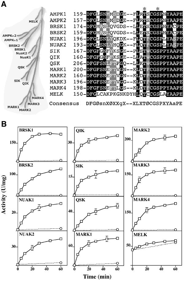
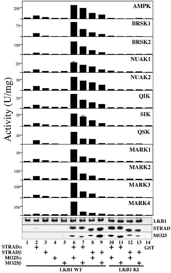
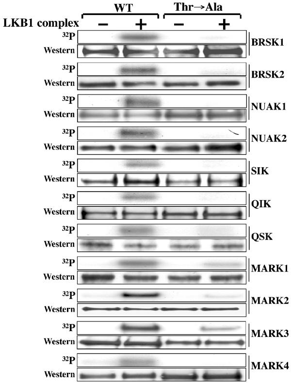
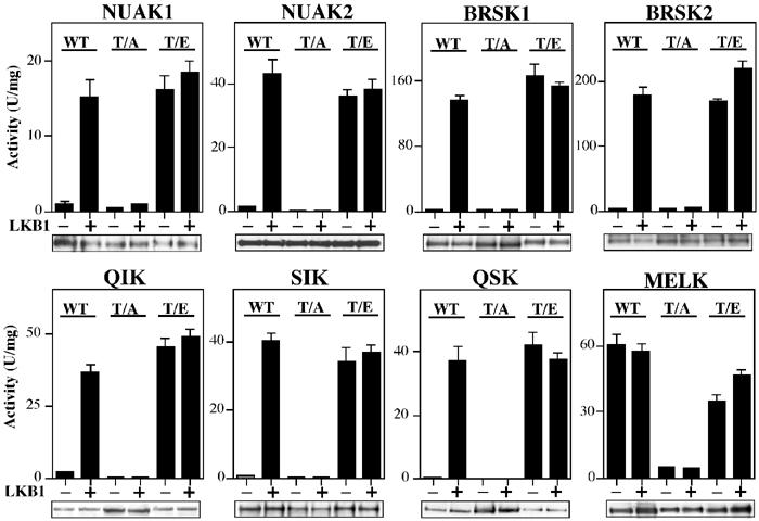
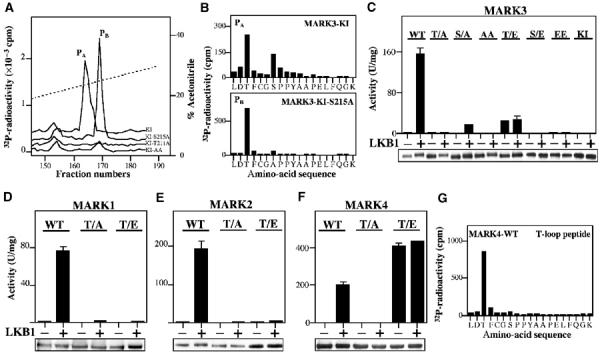
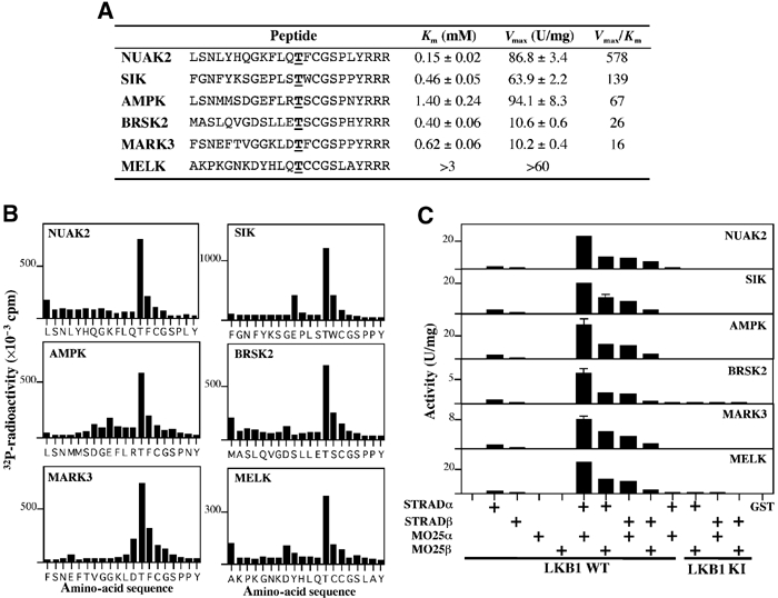
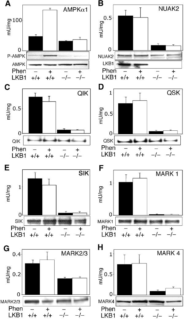
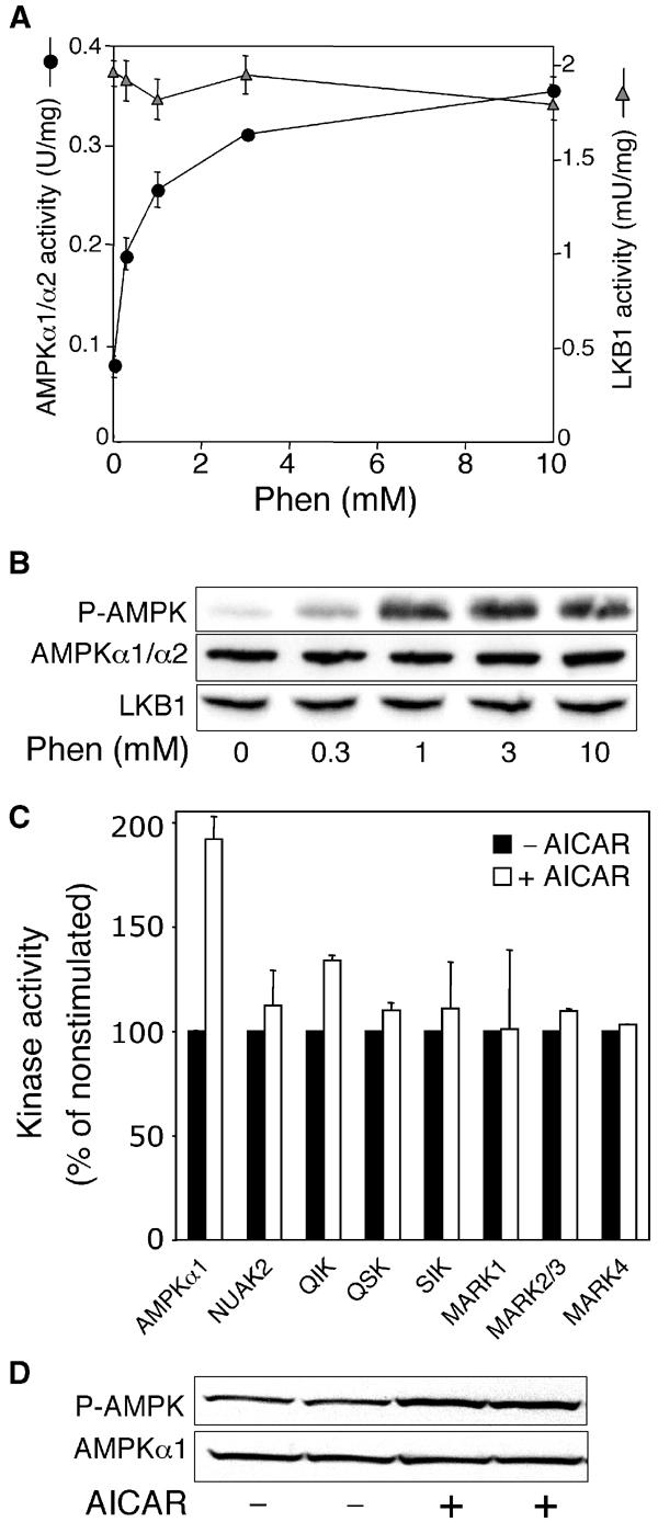
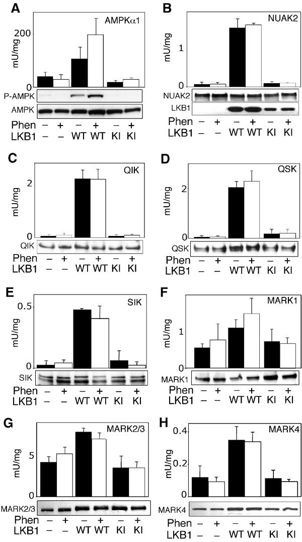
We recently demonstrated that the LKB1 tumour suppressor kinase, in complex with the pseudokinase STRAD and the scaffolding protein MO25, phosphorylates and activates AMP-activated protein kinase (AMPK). A total of 12 human kinases (NUAK1, NUAK2, BRSK1, BRSK2, QIK, QSK, SIK, MARK1, MARK2, MARK3, MARK4 and MELK) are related to AMPK. Here we demonstrate that LKB1 can phosphorylate the T-loop of all the members of this subfamily, apart from MELK, increasing their activity >50-fold. LKB1 catalytic activity and the presence of MO25 and STRAD are required for activation. Mutation of the T-loop Thr phosphorylated by LKB1 to Ala prevented activation, while mutation to glutamate produced active forms of many of the AMPK-related kinases. Activities of endogenous NUAK2, QIK, QSK, SIK, MARK1, MARK2/3 and MARK4 were markedly reduced in LKB1-deficient cells. Neither LKB1 activity nor that of AMPK-related kinases was stimulated by phenformin or AICAR, which activate AMPK. Our results show that LKB1 functions as a master upstream protein kinase, regulating AMPK-related kinases as well as AMPK. Between them, these kinases may mediate the physiological effects of LKB1, including its tumour suppressor function.


Figures

Figure 1
Figure 1
Activation of AMPK-related kinases by LKB1. (A) Dendrogram and T-loop sequences of AMPK subfamily of protein kinases (Manning et al, 2002). The identical residues are shaded black and the conserved residues in grey. The T-loop Thr and Ser are indicated with an asterisk. (B) The indicated AMPK-related kinases were incubated with wild-type LKB1:STRAD:MO25 (open squares) or catalytically inactive LKB1[D194A]:STRAD:MO25 (open circles) complexes in the presence of Mg2+ and ATP. At the indicated times, the activity of the AMPK-related kinases was assayed with the AMARA peptide, and the results are expressed as specific activity. Results shown are means±s.d. of assays carried out in triplicate and representative of two independent experiments. The error bars are only shown when larger than the size of the open squares. The suggested consensus sequence for optimal LKB1 phosphorylation is indicated. Ø represents a large hydrophobic residue; X, any amino acid; s, n, g and a preferences for Ser, Asn, Gly and Ala, respectively.
Figure 2
Figure 2
Efficient activation of AMPK-related kinases by LKB1 requires STRAD and MO25 subunits. The indicated combinations of GST-tagged wild-type LKB1 (WT, lanes 1–9), or catalytically inactive (KI, D194A, lanes 10–13) LKB1, or GST alone (lane 14), FLAG-tagged STRADα or STRADβ, and myc-tagged MO25α or MO25β were coexpressed in HEK-293T cells and purified on glutathione–sepharose. The complexes were tested for their ability to activate the catalytic domain of AMPKα1 or the indicated AMPK-related kinases. The results are expressed as specific activity employing the AMARA peptide as substrate. Results shown are means±s.d. of assays carried out in triplicate and representative of two independent experiments. Samples from each incubation were also analysed by Western blotting and probed using the indicated antibodies (from top to bottom): anti-GST to detect LKB1; anti-FLAG to detect STRADα and STRADβ; and anti-myc to detect MO25α and MO25β. All proteins migrated with the expected mobility, taking into account the epitope tags.
Figure 3
Figure 3
The T-loop Thr is the major site of LKB1 phosphorylation on the AMPK-related kinases. Wild-type (WT) and T-loop Thr to Ala (Thr → Ala) mutants of the indicated GST-AMPK-related kinases were incubated with the LKB1:STRAD:MO25 complex in the presence of Mg2+ and [γ32P]ATP. Phosphorylation of protein substrates was determined by electrophoresis on a polyacrylamide gel and subsequent autoradiography of the Coomassie blue-stained bands corresponding to each substrate. An aliquot of each incubation was also analysed by Western blotting probing with an anti-HA antibody to ensure equal loading of wild-type and mutant AMPK-related kinases (which all possess an HA epitope tag). All proteins migrated with the expected mobility, taking into account the epitope tags. Similar results were obtained in three separate experiments.
Figure 4
Figure 4
Effect of mutation of Thr in the T-loop on activation of AMPK-related kinases by LKB1. The indicated wild-type (WT) AMPK-related kinases or mutants of these enzymes in which the T-loop Thr was changed to either Ala (T/A) or Glu (T/E) were incubated in the absence (−) or presence (+) of wild-type LKB1:STRAD:MO25 in the presence of Mg2+ and ATP. After 30 min, the AMPK-related kinases were assayed with the AMARA peptide, and the results are expressed as specific activity. Results shown are means±s.d. of assays carried out in triplicate and representative of two independent experiments. An aliquot of each incubation was also analysed by Western blotting probing with an anti-HA antibody to ensure equal loading of wild-type and mutant AMPK-related kinases (which all possess an HA tag).
Figure 5
Figure 5
Analysis of phosphorylation and activation of MARK kinases. (A) Catalytically inactive MARK3[D196A] (KI), which cannot autophosphorylate, and the indicated mutants were incubated with the LKB1 complex for 30 min with Mg2+-[γ32P]ATP and separated by electrophoresis on a polyacrylamide gel, which was then autoradiographed. The 32P-labelled MARK3 proteins were digested with trypsin and the resulting 32P-labelled peptides were chromatographed on a C18 column. Fractions containing the 32P-labelled T-loop tryptic peptide (peptides PA and PB) are shown. (B) Peptide PA and PB were subjected to solid-phase sequencing and 32P-radioactivity was measured after each cycle of Edman degradation. In combination with MALDI TOF–TOF mass spectrometry, this enabled the identification of the sites phosphorylated in each peptide. Peptide PA comprise the MARK3 T-loop peptide phosphorylated at Thr211 and Ser215, and Peptides PB comprise the MARK3 T-loop peptide phosphorylated at Thr211. (C–F) The indicated wild-type (WT) or mutant forms of MARK kinases in which the the T-loop Thr or Ser was mutated to either Ala (T/A, S/A) or Glu to (T/E, S/E) were incubated in the absence (−) or presence (+) of wild-type LKB1:STRAD:MO25 in the presence of Mg2+ and ATP. After 30 min, the MARK kinases were assayed using the AMARA peptide, and the results are expressed as specific activity. An aliquot of each incubation was also analysed by Western blotting probing with an anti-HA antibody to ensure equal loading of wild-type and mutant MARK kinases (which all possessed an HA epitope tag). Results shown are average±s.d. of a triplicate assay and are representative of at least two independent experiments. (G) MARK4 was phosphorylated with the LKB1 complex as in (A) and the major 32P-labelled peptide was analysed by solid-phase Edman sequencing as in (B). In combination with MALDI TOF–TOF mass spectrometry (Supplementary Figure 1C), this peptide was shown to comprise the T-loop of MARK4 phosphorylated at only the Thr residue.
Figure 6
Figure 6
Identification of peptide substrates for LKB1. (A) Kinetic analysis of the phosphorylation of the indicated T-loop by the LKB1:STRAD:MO25 complex was performed. The T-loop Thr residue in each peptide is underlined and is in boldface type. Three Arg residues were added to the C-terminus of each T-loop peptide to enable their capture on phosphocellulose p81 paper. Km and Vmax values were determined from nonlinear regression. (B) An aliquot of each peptide phosphorylated by the LKB1 complex was subjected to solid-phase Edman sequencing and 32P-radioactivity was measured after each cycle of Edman degradation. A small proportion of each peptide can become coupled to the Sequelon arylamine membrane through acidic internal Asp and Glu residues rather than their C-terminal carboxyl group. This accounts for the apparent small releases of 32P-radiactivity that are observed at some Asp and Glu residues. (C) Same combinations of GST-tagged wild-type LKB1 (WT, lanes 1–9), or catalytically inactive (D194A, lanes 10–13) LKB1, or GST alone (lane 14), FLAG-tagged STRADα or STRADβ, and myc-tagged MO25α or MO25β employed in Figure 2 were tested for their ability to phosphorylate the indicated peptides (peptide concentration 200 μM). The results are expressed as the peptide kinase activity generated per mg of LKB1:STRAD:MO25 added to the assay. Results shown are average±s.d. of three assays and are representative of at least two independent experiments.
Figure 7
Figure 7
Activity of AMPK-related kinases in LKB1+/+ and LKB1−/− MEFs. LKB1+/+ or LKB1−/− MEFs were either left untreated (black bars) or stimulated with 10 mM phenformin (Phen, white bars) for 1 h. AMPKα1 and AMPK-related kinases were immunoprecipitated from the cell lysates, and in vitro kinase activity towards the AMARA peptide was measured as described in Materials and methods. To confirm equal expression of the kinases in each sample, cell lysates (AMPKα1, NUAK2, MARK2/3 and MARK4) or immunoprecipitated proteins (QIK, QSK, SIK and MARK1) were subjected to SDS–PAGE and Western blot analysis. All AMPK-related kinases migrated with the expected molecular mass. In the case of AMPKα1, cell lysates were immunoblotted with a phospho-Thr172 antibody (P-AMPK) that recognises the phosphorylated T-loop. A control immunoblot of LKB1 levels in cell lysates is also included in panel B. Results shown are average±s.d. of 2–4 assays and are representative of at least two independent experiments.
Figure 8
Figure 8
AICAR does not activate AMPK-related kinases. (A) LKB1+/+ MEFs were either left untreated or stimulated with the indicated concentrations of phenformin (Phen) for 1 h. AMPKα1/α2 and LKB1 were immunoprecipitated from the cell lysates, and in vitro kinase activity towards the AMARA and LKBtide peptides, respectively, was measured as described in Materials and methods. Results shown are average±s.d. of a triplicate assay and are representative of two independent experiments. (B) To confirm equal expression of the kinases in each sample, cell lysates were subjected to SDS–PAGE and Western blot analysis with the indicated antibodies. The phospho-Thr172 antibody (P-AMPK) recognises the phosphorylated T-loop of AMPKα1. (C) LKB1+/+ MEFs were either left untreated (black bars) or stimulated with 2 mM AICAR (white bars) for 1 h. AMPKα1 and the indicated AMPK-related kinases were immunoprecipitated from the cell lysates, and in vitro kinase activity towards the AMARA peptide was measured as described in Materials and methods. Results are presented as % relative to the activity observed in nonstimulated cells, and are averages±s.e.m. of two independent experiments. 100% corresponds to the following absolute activities: AMPKα1, 135 mU/mg; NUAK2, 0.25 mU/mg; QIK, 0.76 mU/mg; QSK, 1.9 mU/mg; SIK, 2.73 mU/mg; MARK1, 0.14 mU/mg; MARK2/3, 3.5 mU/mg; and MARK4, 1.6 mU/mg. (D) Immunoblotting of AMPK was performed as in (B).
Figure 9
Figure 9
Activity of AMPK-related kinases in HeLa cells. Control HeLa cells lacking LKB1 expression (−), or HeLa cells stably expressing wild-type LKB1 (WT) or kinase inactive LKB1 (KI), were either left untreated (black bars) or stimulated with 10 mM phenformin (Phen, white bars) for 1 h. AMPKα1 and AMPK-related kinases were immunoprecipitated from the cell lysates, and in vitro kinase activity towards the AMARA peptide was measured as described in Materials and methods. To confirm equal expression of the kinases in each sample, cell lysates (AMPKα1, MARK2/3 and MARK4) or immunoprecipitated proteins (NUAK2, QIK, QSK, SIK and MARK1) were subjected to SDS–PAGE and Western immunoblot analysis. All AMPK-related kinases migrated with the expected molecular mass. In the case of AMPKα1, cell lysates were immunoblotted with a phospho-Thr172 antibody (P-AMPK) that recognises the phosphorylated T-loop. A control immunoblot of LKB1 levels in cell lysates is also included in panel B. Results shown are average±s.d. of 2–4 assays and are representative of at least two independent experiments.

Similar articles

Cited by

References

    1. Alessi DR (2001) Discovery of PDK1, one of the missing links in insulin signal transduction. Biochem Soc Trans 29: 1–14 - PubMed
    1. Baas AF, Boudeau J, Sapkota GP, Smit L, Medema R, Morrice NA, Alessi DR, Clevers HC (2003) Activation of the tumour suppressor kinase LKB1 by the STE20-like pseudokinase STRAD. EMBO J 22: 3062–3072 - PMC - PubMed
    1. Boudeau J, Baas AF, Deak M, Morrice NA, Kieloch A, Schutkowski M, Prescott AR, Clevers HC, Alessi DR (2003a) MO25alpha/beta interact with STRADalpha/beta enhancing their ability to bind, activate and localize LKB1 in the cytoplasm. EMBO J 22: 5102–5114 - PMC - PubMed
    1. Boudeau J, Deak M, Lawlor MA, Morrice NA, Alessi DR (2003b) Heat-shock protein 90 and Cdc37 interact with LKB1 and regulate its stability. Biochem J 370: 849–857 - PMC - PubMed
    1. Boudeau J, Sapkota G, Alessi DR (2003c) LKB1, a protein kinase regulating cell proliferation and polarity. FEBS Lett 546: 159–165 - PubMed

Publication types

MeSH terms