. 2004 Mar 10;23(5):1000-7.
doi: 10.1038/sj.emboj.7600115. Epub 2004 Feb 19.

Structure of the La motif: a winged helix domain mediates RNA binding via a conserved aromatic patch

Affiliations

Structure of the La motif: a winged helix domain mediates RNA binding via a conserved aromatic patch

Gang Dong et al. EMBO J. .

Abstract

The La protein is a ubiquitous nuclear phosphoprotein that recognizes the 3' uridylates found in all newly synthesized RNA polymerase III transcripts. La binding stabilizes these transcripts from exonucleases and may also assist their folding. Here we present the first structural insights into how the La protein specifically interacts with its RNA substrates. The most conserved region of the La protein is the La motif, a domain also found in several other RNA-binding proteins. We have determined the structure of the La motif from the Trypanosoma brucei La protein to 1.6 A resolution (PDB code 1S29). The La motif adopts a winged helix-turn-helix architecture that has a highly conserved patch of mainly aromatic surface residues. Mutagenesis experiments support a critical role for this patch in RNA binding and show that it partly determines binding specificity for RNAs ending in 3' hydroxyl, a defining characteristic of the La protein. These findings reveal that the La motif is essential for high-affinity binding and also contributes to specificity.


Figures

Figure 1
Figure 1
(A) Ribbon views of the LM of T. brucei La. (B) Ribbon view of a wHTH motif as observed in the Z-DNA-binding domain of the adenosine deaminase ADAR1 (PDB accession number 1QBJ). The topology of the LM is similar to that of the wHTH motif of some transcription factors. MOLSCRIPT (Kraulis, 1991) was used to prepare this figure.
Figure 2
Figure 2
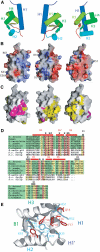
The orientations of the LM in (A–C) are the same. The three orientations are related by a rotation about an axis along the length of the page. (A) Ribbon representation of the LM with cylinders representing all helices but H1′. Helix H1′ is rendered as a squiggle. (B) Qualitative map of the electrostatic potential at the LM surface. The left-most view shows the positively charged surface adjoining the conserved aromatic patch (marked by D27). (C) Views of the LM are as in (B), but pink surfaces indicate residues that are invariant in La proteins. The conserved aromatic patch is visible in the left-most image. Yellow denotes less strictly conserved residues. Asterisks mark conserved surfaces that were mutated. (D) An alignment of LM sequences. Sequences from true La proteins, which bind nascent RNA polymerase III RNAs, are boxed in green. The Caenorhabditis elegans protein has not been biochemically characterized. Sequences for Slf1p, Sro9p, and p43, which contain an LM but are not La proteins, are also shown. As in (C), residues that are invariant in the true La proteins are boxed in pink and conserved residues are boxed in yellow. Red bars indicate α-helices, and striped black bars indicate β-strands. Stars indicate those residues that form an extended conserved patch on the surface of the LM. The patch is essential for RNA binding and recognition. (E) A rendering of the aromatic patch conserved in all LM proteins. Mutating the residues depicted in red eliminated La binding to pre-tRNA. Mutating residues D27, N23, and R52 to alanine had modest effects on binding affinity, and D27A had a reduced specificity for a hydroxyl over a phosphate group at the RNA 3′ terminus. MOLSCRIPT (Kraulis, 1991) was used to prepare (A, E) and Grasp (Nicholls et al, 1991) was used to prepare (B, C).
Figure 3
Figure 3
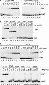
Binding of wild-type and mutant La proteins to RNA. (A) 32P-labeled yeast pre-tRNACCGArg (left panel) or pre-tRNACCGArg lacking the 9 nt 3′ trailer (right panel) was incubated without protein (lane 1) or with the truncated T. brucei La protein (LM+RRM; amino acids 1–192) at the indicated protein concentrations. The pre-tRNA concentration in the reaction was 0.15 nM. Naked RNAs and RNPs were separated in native gels. (B) 32P-labeled yeast pre-tRNACCGArg (final concentration 0.15 nM) was incubated with either no protein (lane 1) or the indicated concentrations of full-length La (lanes 2–4), La 1–192 (lanes 5–8), the isolated LM (amino acids 1–89; lanes 9–11), the RRM (residues 94–192; lanes 12–14), or a 1:1 mixture of the LM and RRM (lanes 15–17). Naked RNAs and RNPs were separated by native gel electrophoresis. (C) Binding of La 1–192 (left panel) and the D27A mutant (right panel) to pre-tRNACCGArg was performed as described in (A). Protein concentrations in the reactions are indicated above the lanes. In lanes 1 and 10, no protein was added. We quantitated the difference in binding affinity of La 1–192 and the D27A mutant by performing multiple experiments. These revealed that La 1–192 bound the RNA with a Kd of 4.1±0.7 nM, while the D27A mutant bound the RNA with a Kd of 14.6±2.9 nM, an approximately 3.6-fold decrease in affinity. (D) pre-tRNACCGArg was subjected to gel shift analysis using either the wild-type La 1–192 (lanes 2–4) or the mutants Q14A/E16A (lanes 5–7), F29A (lanes 8–10), F50A (lanes 11–13), F17A (lanes 14–16), and Y18A (lanes 17–19). Protein concentrations in each lane are given in nM. Lanes 1 and 20, no protein.
Figure 4
Figure 4
Mutating D27 reduces the specificity of La for RNAs ending in a 3′ hydroxyl. (A) La 1–192 (4.8 nM) was mixed with 32P-labeled pre-tRNACCGArg (0.15 nM) in the presence of a 0- to 10 000-fold molar excess of unlabeled pre-tRNACCGArg terminating in either UUUp (left panel) or UUUOH (right panel). Naked RNAs and RNPs were separated by native gel electrophoresis. The small differences in the fraction of RNA bound with 4.8 nM protein between this experiment and Figure 3C, lane 5, reflect experimental variation. Lane 1, no added protein. (B, C) The N23A mutant (B) or D27A mutant protein (C) was mixed with 32P-labeled pre-tRNACCGArg (0.15 nM) in the presence of the indicated molar excess of unlabeled pre-tRNACCGArg as described in (A). The concentration of protein used in the reactions was 19.2 nM. Lane 1, no added protein. (D) Data from the competition titrations for La 1–192 and the D27A mutant are plotted as the fraction of labeled probe bound at each concentration of competitor RNA. Each data point represents the mean from at least three experiments. RNA concentrations are given in nM.

References

    1. Aigner S, Postberg J, Lipps HJ, Cech TR (2003) The Euplotes La motif protein p43 has properties of a telomerase-specific subunit. Biochem 42: 5736–5747 - PubMed
    1. Allers J, Shamoo Y (2001) Structure-based analysis of protein–RNA interactions using the program ENTANGLE. J Mol Biol 311: 75–86 - PubMed
    1. Alspaugh MA, Tan EM (1975) Antibodies to cellular antigens in Sjogren's syndrome. J Clin Invest 55: 1067–1073 - PMC - PubMed
    1. Antson A (2000) Single stranded RNA binding proteins. Curr Opin Struct Biol 10: 87–94 - PubMed
    1. Bai C, Li Z, Tolias PP (1994) Developmental characterization of a Drosophila RNA-binding protein homologous to the human systemic lupus erythematosus-associated La/SS-B autoantigen. Mol Biol Cell 14: 5123–5129 - PMC - PubMed

Publication types

MeSH terms