. 2004 Jan-Feb;256-257(1-2):243-56.
doi: 10.1023/b:mcbi.0000009872.35940.7d.

Nucleotide-gated KATP channels integrated with creatine and adenylate kinases: amplification, tuning and sensing of energetic signals in the compartmentalized cellular environment

Affiliations

Nucleotide-gated KATP channels integrated with creatine and adenylate kinases: amplification, tuning and sensing of energetic signals in the compartmentalized cellular environment

Vitaliy A Selivanov et al. Mol Cell Biochem. 2004 Jan-Feb.

Abstract

Transmission of energetic signals to membrane sensors, such as the ATP-sensitive K+ (KATP) channel, is vital for cellular adaptation to stress. Yet, cell compartmentation implies diffusional hindrances that hamper direct reception of cytosolic energetic signals. With high intracellular ATP levels, KATP channels may sense not bulk cytosolic, but rather local submembrane nucleotide concentrations set by membrane ATPases and phosphotransfer enzymes. Here, we analyzed the role of adenylate kinase and creatine kinase phosphotransfer reactions in energetic signal transmission over the strong diffusional barrier in the submembrane compartment, and translation of such signals into a nucleotide response detectable by KATP channels. Facilitated diffusion provided by creatine kinase and adenylate kinase phosphotransfer dissipated nucleotide gradients imposed by membrane ATPases, and shunted diffusional restrictions. Energetic signals, simulated as deviation of bulk ATP from its basal level, were amplified into an augmented nucleotide response in the submembrane space due to failure under stress of creatine kinase to facilitate nucleotide diffusion. Tuning of creatine kinase-dependent amplification of the nucleotide response was provided by adenylate kinase capable of adjusting the ATP/ADP ratio in the submembrane compartment securing adequate KATP channel response in accord with cellular metabolic demand. Thus, complementation between creatine kinase and adenylate kinase systems, here predicted by modeling and further supported experimentally, provides a mechanistic basis for metabolic sensor function governed by alterations in intracellular phosphotransfer fluxes.


Figures

Fig. 1
Fig. 1
Stoicheometry and allosteric nucleotide-dependent gating of KATP channels. (A) The KATP channel is an octameric complex composed of four pore-forming Kir6.2 subunits and four associated regulatory SUR subunits. Kir6.2 is formed by two transmembrane domains flanking a pore region. SUR is characterized by two cytosolic nucleotide-binding domains (NBD1 and NBD2) which contain highly-conserved Walker A and Walker B motifs as well as a linker (L) region. Interaction of ATP (green triangle) with Kir6.2 induces pore closure. Upper channel record demonstrates ATP-induced KATP channel inhibition. MgADP (blue triangle) at NBD2 antagonizes ATP-induced pore inhibition, with an apparent requirement of ATP at NBD1. Lower channel record demonstrates MgADP-induced reduction of ATP-induced KATP channel inhibition leading to channel opening. (B) MgADP antagonizes ATP-induced inhibition of cardiac KATP channels. In excised patches, the ATP sensitivity of KATP channels was defined by an IC50 of 27 ± 5 mM in the absence (triangles) vs. 270 ± 19 μM in the presence (circles) of 100 μM ADP. Relative channel activity (curves) constructed based on an allosteric model of nucleotide-dependent KATP channel gating (C; see Appendix), and expressed as a probability for the channel to be in an open state (columns T0-Tl. In the absence of ADP, channels adopt the highest sensitivity to ATP (row Do) defined solely by the microscopic dissociation constant k0 = 45 μM (curve 1). At saturating ADP concentrations, KATP channels convert to channel species with the lowest ATP sensitivity (row D4) defined solely by k1 = 450 μM (curve 4). kADP (12.5 μM) was determined at different concentrations of ADP (curve 1: 0; 2: 10; 3: 50; 4: 100; 5: 500; 6: 1000 μM ADP).
Fig. 2
Fig. 2
KATP channels communicate with intracellular phosphotransfer systems. To maintain the relative integrity of the cellular infrastructure, KATP channel activity was recorded in the open-cell attached patch mode in cardiac cells from wild-type and M-CK or AK1-deficient mice (see [22, 34]). (A) CrP-dependent regulation of KATP channels is lost in M-CK-knockout cardiac cells. While in the wild-type, CrP enhanced KATP channel inhibition by 100 μM ATP (upper trace), in M-CK knockouts (lower trace) the creatine kinasc substrate was deprived of a significant effect. Temperature was 31°C. Adapted from [34]. (B) AMP-dependent regulation of KATP channels is lost in AK1 -knockout cardiac cells. KATP channel recordings demonstrate activation of ATP-inhibited channels following application of AMP, an adenylate kinase substrate (upper trace), but absence of AMP effect in cardiomyocytes from AK 1 -knockout hearts. Measurements were at room temperature. Adapted from [11].
Fig. 3
Fig. 3
Adenylate kinase diminishes nucleotide gradients between cellular compartments. (A) ATP-dependence of relative channel activity constructed based on the allosteric model of KATP channel gating (k0 = 10 μM, k1 = 450 μM, kADP = 12.5 μM; see Appendix) at (from left to right) 0,0.01, 0.05, 0.1, 0.5 and 1.0 mM MgADP, with further addition of ADP not shifting the curve. Vertical bar indicates concentration of ATP (3 mM), at which at least 1 % of KATP channel population could be opened at saturated ADP level. (B) The model used to estimate diffusion coefficient for adenine nucleotides in submembrane space (Eqs 1-6). ATPase in submembrane space creates nucleotide gradients modified by the AK reaction operating at a presumed equilibrium in both compartments. Gradients cause nucleotide diffusion fluxes (horizontal arrows) over diffusion barrier according to Fick's law. The diffusion coefficient 1.5 10-11 cm2/s was calculated (Eqs 1, 2 and 4) based on the assumption that at 7 mM of total nucleotides, 6.0 mM of bulk ATP ([ATP]b) and a membrane ATPase activity of a working heart (4.7 10-6 μmol/cm2/s), submembrane ATP ([ATP]m) levels must drop to 3 mM in order to secure minimum KATP channel activity (1% of the channel population) sufficient for changes in cardiac membrane excitability. (C) Differences in ATP, ADP and AMP concentrations (ΔATP, ΔADP and ΔAMP) between the bulk and submembrane space estimated using Eq. 6 as function of bulk ATP. (D) Expected submembrane nucleotide concentration corresponding to nucleotide concentration gradients presented in (C). (E) Differences in nucleotide concentrations between the bulk and sub-membrane space computed at various membrane ATPase activities; (-AK) indicates curves constructed assuming absence of AK activity.
Fig. 4
Fig. 4
Creatine kinase as a submembrane amplifier of cytosolic changes in ATP and ADP. (A) Model of CrP/CK - facilitated diffusion for adenine nucleotide (Eqs 7-13). ATPase and creatine kinase reactions (assumed at equilibrium) in the submembrane space create nucleotide and creatine gradients that cause diffusional fluxes (horizontal arrows) according to Fick's law. (B) Profiles of ΔATP and submembrane concentration of creatine phosphate ([CrP]m) constructed based on Eq. 13 as a function of bulk ATP ([ATP]b), at 7 mM total nucleotide content, 40 mM total Cr/CrP level, ATPase flux 4.7 10-6umol/cm2/s, diffusion coefficient 2.25 10-11 cm2/s, and the equilibrium constant for the CK reaction KCK = 160. (C) The sub-membrane concentration of ATP and ADP at the parameters used in (B). (D) Differences in nucleotide concentrations between bulk and sub-membrane space at various membrane ATPase activities, [ATP]b = 6.99 mM in the absence (-CK) or presence (+CK) of CK activity.
Fig. 5
Fig. 5
Adenine nucleotide diffusion facilitated by the CrP/CK system in the presence of AK.
Fig. 6
Fig. 6
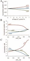
Regulation of adenine nucleotides by coactivc creatine and adenylate kinases. (A) Differences in nucleotide concentrations between hulk and sub-membrane space at various membrane ATPase activities obtained by resolving the system of Eqs 2,4,5,8,9, 11,12, and 14 with corresponding parameters as in Figs 3E and 4D. (B) Differences in nucleotide concentrations between bulk and submembrane compartments at membrane ATPase activity = 4.7 10-6 μmol/cm2/s and various [ATP]b. (C) Submembrane nucleotidc concentrations that correspond to ΔATP, ΔADP, and ΔAMP shown in (B).
Fig. 7
Fig. 7
Amplification and tuning of energetic signals by CK and AK. Energetic signal was simulated as a slow-rate, gaussian, dip in [ATP]b. (A) CK amplifies a 0.5 mM drop in [ATP]b into a ~ 6 time higher change in [ATP]m corresponding to a significant response in the [ATP]m/[ADP]m ratio (upper row). AK provides a ~ 2 times attenuation of [ATP]m in response to the 0.5 mM drop in [ATP]b, insufficient to induce significant changes in the [ATP]m/[ADP]m ratio (middle row). Coactive AK and CK provide significant amplification of a [ATP]b signal, yet doubling in [ATP]b signals (0.5, 1, 2, and 4 mM dips) transmit into an amplified and modulated (2.6, 3.7, 5.1, and 6.6 mM, respectively) response in [ATP]m and a cut-off of [ATP]m/[ADP]m ratio (lower row). Note that higher signals undergo a lower amplification, an effect enhanced by AK. (B) Effectiveness of signal transmission represented as a derivative of changes in [ATP]m over [ATP]b for CK alone and co-active CK and AK systems. Dotted line corresponds to a passive signal response in the absence of systems catalyzing phosphotransfer reactions. (C) In rat cardiomyocytes, in the open cell-attached mode of the patch-clamp technique, ATP-induced KATP channel inhibition was antagonized by activation of membrane Na+/K+ ATPase following application of 40 mM of NaCl. Ouabain, an inhibitor of the Na+/K+ pump, reversed ATP-induced channel closure (upper trace). Di(adenosine-5′) pentaphosphate (P1,P5), a selective AK inhibitor, produced additional opening of KATP channels under active membrane Na+/K+ ATPase, preventing AK scavenging of ADP and support of ATP levels in the submembrane compartment. (D) Concentration-response curves defining ATP-induced KATP channel inhibition measured from guinea-pig cardiomyocytes in open cell-attached mode in the absence (JCrP = 0, open triangles) and in the presence of 1 mM CrP (JCrp >> 0, closed triangles). Solid curves were constructed based on the allosteric model of channel regulation (with k0 = 10 μM, k1 = 450 μM, kADP = 12.5 μM), in conjunction with nucleotide diffusion, JATPase and creatine kinase (JCK) fluxes (see [34]). Activation of CK flux produced a significant left-shift of ATP-induced KATP channel inhibition from IC50 = 270 ± 2 μM at JCrp = 0 to IC50 = 7 ± 1 μM at JCrP >> 0. Following changes in [ATP]b, without changes in JCrP, KATP channels remain closed in accord with a higher ATP sensitivity (red arrow). Assuming that the proposed shift in [ATP]b corresponds to complete suppression of JCrP, KATP channel activity can be determined as a transition to the curve defining lower ATP sensitivity (blue arrow) resulting in amplified changes in open channel probability. (E) Transmission of an amplified energetic signal from the bulk space to the KATP channel site by altered CK flux was assessed in mice cardiomyocytes, under active membrane Na+/K+ ATPase, in open cell-attached patches. S1, S2 and S3 solutions, with compositions presented in the table, simulated different values for CK fluxes. In the Table, applied concentrations are in normal typeface, whereas actual concentrations defined according to the reaction equilibrium (Eq. 9) are bold in parentheses. In order to secure reaction equilibrium in the bulk space, all solutions were supplemented with exogenous CK (170-200 U/ml) in addition to intracellular CK. Vigorous KATP channel activity was readily inhibited by high JCrP simuxlated by S1 at 0.3 mM [ATP]b. Low JCrP (solution S2) led to KATP channel opening amplifying minor changes in [ATP]b and [ADP]b within the submembrane compartment. The amount of CrP used in S2 was sufficient to reverse KATP channel inhibition in the absence of Cr (solution S3). Measurements were performed at 60 mV, and 31 ± 1°C.
Fig. 8
Fig. 8
KATP channels as sensors of metabolic signals transmitted over diffusion barrier by the creatine and adenylate kinases. (A) Solid curve depicts regulation of KATP channels by submembrane nucleotides, calculated based on the allosteric model (see Appendix) at 7 mM of total nucleotide and various [ATP]b. In the absence of stress co-active CK and AK essentially void nucleotide gradients, equalizing [ATP]b and [ATP]m, and keeping KATP channels closed. A stress-induced 1 mM drop of [ATP]b (ΔATPb, small bar), would be amplified into a dramatic drop in [ATP]m (ΔATPm, large bar) permitting opening of > 1% of KATP channels (intersection of the right edge of the bar for ΔATPm with solid curve). (B) Threshold for significant shortening of cardiac action potential represented as a bar filling the 1-5% range of open KATP channels. Channel activity calculated based on the allosteric model of channel regulation by submembrane nucleotides at various [ATP]b and different membrane ATPase activities. In the absence of adenylate kinase (-AK) the extremely steep nucleotide-dependent channel regulation (dashed line) would expose cells to excessive KATP current at relatively modest changes in [ATP]b. (C) AK system may favor KATP channel openings at active bulk ATPase, i.e. following vigorous mitochondria uncoupling, when [ATP]m > [ATP]b. Such conditions require the support of [ATP]m which was simulated through diffusional flux of ATP (J0ATP) and CrP (J0CrP) into the submembrane space from a third compartment with a constant level of [ATP]0 = 0.5 mM and [CrP]0 = 1 mM. Thus, at steady state: J0ATP+J0CrP=JAMP+JATP+JCrP, where J0ATP=DSΔX([ATP]0[ATP]m), J0CrP=DSΔX([CrP]0[CrP]m) whereas other fluxes defined from Eqs 2 and 8. The relation between fluxes (Eq. 15) is similar to Eq. 14, ignoring submembrane ATPase activity (JATPase). The percentage of opened KATP channels was estimated using the allosteric model for channel regulation by submembrane nucleotide levels, that were calculated for co-active CK and AK activities (+AK, Eqs 2, 4, 5, 8, 9, 11, 12, and 15), vs. CK alone (-AK, Eqs 8, 9, 11, 12, and 15) at various bulk ATPase-induced drop of [ATP]b. (D) Activity of KATP channels measured in cardiomyocytes isolated from wile-type (upper trace) and AK1-deficient (AK1-KO, lower trace) mice under simulated conditions described above (C). To preserve intracellular catalytic systems recording were performed in the open cell-attached mode at 30°C [see 11]. In wild-type, P1,P5 inhibited adenylate kinase activity and effectively inhibited DNP-induced KATP channel openings ([ATP]0 = 0.5 mM, [CrP]0 = 1 mM) indicating involvement of the AK system in channel openings (note, that membrane ATPase activity in resting cardiomyocytes, as in such experimental conditions, is much lower in comparison with cells in working heart). In AK1-knockout cardiomyocytes, the KATP channel response to DNP-induced mitochondrial ATPase activation was blunted and sensitive to oligomycin, but insensitive to P1,P5 ([ATP]0 = 0.25 mM, [CrP]0 = 1 mM; [adapted from 11]).

Similar articles

Cited by

References

    1. Aliev MK, Saks VA. Compartmentalized energy transfer in cardiomyocytes: Use of mathematical modeling for analysis of in vivo regulation of respiration. Biophys J. 1997;73:428–445. - PMC - PubMed
    1. O'Rourke B, Ramza BM, Marban E. Oscillations of membrane current and excitability driven by metabolic oscillations in heart cells. Science. 1994;265:962–966. - PubMed
    1. Ventura-Clapier R, Kuznetsov A, Veksler V, Boehm E, Anflous K. Functional coupling of creatine kinases in muscles: Species and tissue specificity. Mol Cell Biochem. 1998;184:231–247. - PubMed
    1. Hardie DG, Carling D, Carlson M. The AMP-activated/SNFl protein kinase subfamily: Metabolic sensors of the eukaryotic cell? Annu Rev Biochem. 1998;67:821–855. - PubMed
    1. Janssen E, Dzeja PP, Oerlemans F, Simonetti AW, Heerschap A, de Haan A, Rush PS, Terjung RR, Wieringa B, Terzic A. Adenylate kinase 1 gene deletion disrupts muscle energetic economy despite metabolic rearrangement. EMBO J. 2000;19:6371–6381. - PMC - PubMed

Publication types

LinkOut - more resources