

The effects of corneal parameters on the assessment of endothelial cell density in the elderly eye
- PMID: 14977761
- PMCID: PMC1772027
- DOI: 10.1136/bjo.2003.019315
The effects of corneal parameters on the assessment of endothelial cell density in the elderly eye
Abstract
Background: The possible impact of corneal thickness, curvature, and size on the measurement of endothelial cell density (ECD) has largely been ignored in the normal eye. The aim of this study was to investigate the possible impact of the main corneal parameters on the analysis of ECD values at the central, superior, and temporal parts of the corneal surface.
Methods: All 75 participants (52 females, 23 males) were assessed as part of a pre-cataract surgery investigation. The mean age was 75.7 (SD 10.9) years. Confocal microscopy was used to measure ECD and the percentage of six sided cells at the central, superior, and temporal parts of the cornea. The Orbscan II topography system was used to measure corneal thickness, topography, and horizontal corneal diameter.
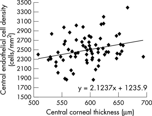
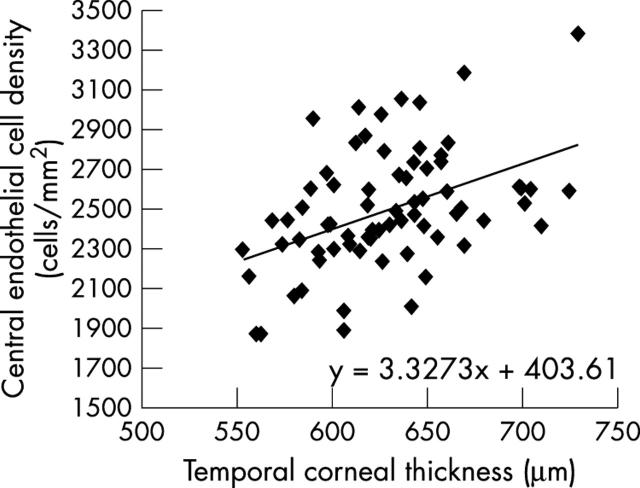
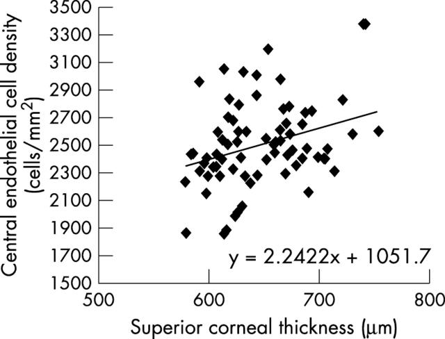
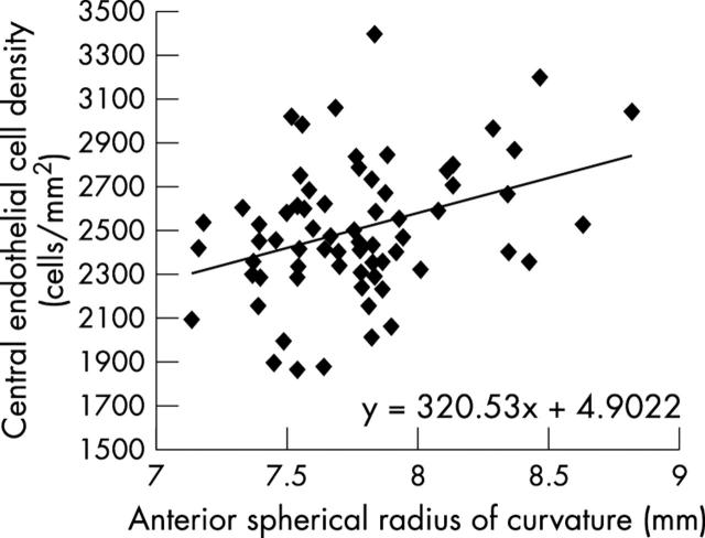
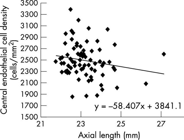
Results: The mean central ECD measured was 2488 (SD 301) cells/mm(2), compared with 2525 (SD 505) cells/mm(2) in the temporal cornea and 2639 (SD 398) cells/mm(2) in the superior cornea. The regional differences in ECD were not significant (p>0.14). The central ECD was significantly correlated to the central (mean 0.593 (SD 0.039) mm, p = 0.021) as well as the temporal (0.628 (SD 0.039) mm, p<0.001) and the superior corneal thickness (SD 0.644 (SD 0.048) mm, p = 0.018). The mean corneal curvature at the centre (7.7 (SD 0.34) mm, p = 0.002) as well as 3 and 5 mm from the apex was significantly related to ECD (p = 0.008 and p = 0.009, respectively).
Conclusions: The study suggests that in an older population, lower ECD values would be expected in thinner and/or steeper corneas.
Figures
References
-
- Laule A, Cable MK, Hoffman CE, et al. Endothelial cell population changes of human cornea during life. Arch Ophthalmol 1978;96:2031–5. - PubMed
-
- Murphy C, Alvarado J, Juster R, et al. Prenatal and postnatal cellularity of the human corneal endothelium. Invest Ophthalmol Vis Sci 1984;25:312–22. - PubMed
-
- Hiles D, Biglan A, Fetherolf E. Central corneal endothelial cell count in children. Am Intr-Ocular Implant Soc J 1979;5:292–300. - PubMed
-
- Hiles D. Discussion to ‘normal endothelial cell count range’. Ophthalmology 1980;87:865–6. - PubMed
-
- Speedwell L, Novakovic P, Sherrard ES, et al. The infant corneal endothelium. Arch Ophthalmol 1988;106:771–5. - PubMed
MeSH terms
LinkOut - more resources
Full Text Sources
Medical
Miscellaneous