. 2004 Mar;72(3):1358-63.
doi: 10.1128/IAI.72.3.1358-1363.2004.

Effect of neutralizing transforming growth factor beta1 on the immune response against Mycobacterium tuberculosis in guinea pigs

Affiliations

Effect of neutralizing transforming growth factor beta1 on the immune response against Mycobacterium tuberculosis in guinea pigs

Shannon Sedberry Allen et al. Infect Immun. 2004 Mar.

Abstract

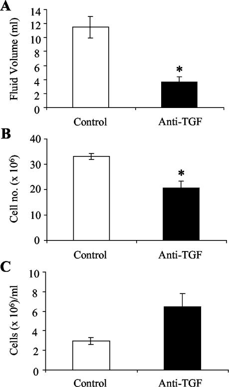
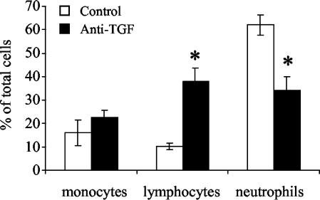
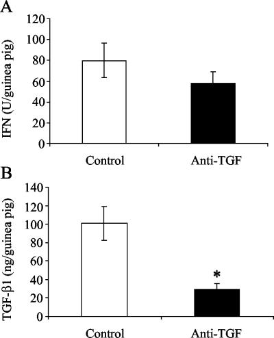
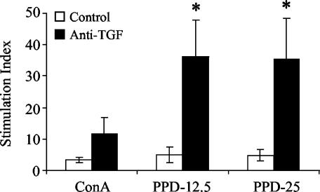
Transforming growth factor beta (TGF-beta) is a cytokine which has been shown to suppress the antimycobacterial immune responses of humans and experimental animals. In this study, the contributions of TGF-beta to cytokine production in vivo were investigated by using the established guinea pig model of tuberculous pleurisy. Mycobacterium bovis BCG-vaccinated guinea pigs were injected intrapleurally with heat-killed virulent Mycobacterium tuberculosis. Eight days following induction of an antigen-specific pleural effusion, guinea pigs were injected intrapleurally with anti-TGF-beta1 or isotype control antibody. The following day, pleural exudates were removed, and the fluid volume and characteristics of the infiltrating cells were determined. Pleural fluid was analyzed for total interferon (IFN) and tumor necrosis factor (TNF) protein levels by using appropriate bioassays. RNA from pleural effusion cells was examined to determine TGF-beta1, TNF-alpha, IFN-gamma, and interleukin-8 mRNA levels by using real-time PCR. Proliferative responses of pleural effusion lymphocytes were examined in response to concanavalin A and purified protein derivative (PPD) in vitro. Treatment with anti-TGF-beta1 resulted in decreased pleural fluid volume and decreased cell numbers in the pleural space along with an increased percentage of lymphocytes and a decreased percentage of neutrophils. The bioactive TNF protein levels in pleural fluid were increased in guinea pigs treated with anti-TGF-beta1, while the bioactive IFN protein concentrations were not altered. Expression of TGF-beta1 and TNF-alpha mRNA was significantly increased following TGF-beta1 neutralization. Finally, PPD-induced proliferative responses of pleural cells from anti-TGF-beta1-treated animals were significantly enhanced. Thus, TGF-beta1 may be involved in the resolution of this local, mycobacterial antigen-specific inflammatory response.


Figures

FIG. 1.
FIG. 1.
Accumulation of fluid and cells in pleural exudates of guinea pigs with tuberculous pleurisy. Fluid accumulation (A), cell accumulation (B), and cell concentration (C) were measured in control pleuritic guinea pigs and guinea pigs receiving experimental injections of anti-TGF-β1. Cells were stained with trypan blue and counted with a hemocytometer. The bars and error bars indicate averages and standard errors of the means for three of four animals. An asterisk indicates that there is significant difference between the number of cells or fluid volume and the number of cells or fluid volume in control guinea pigs.
FIG. 2.
FIG. 2.
Differential counts of pleural exudate cells from pleuritic control and anti-TGF-β1-treated guinea pigs. Cells were stained with Diff-Quik as described in Materials and Methods, and cell morphology was used to determine which cell types were present in the pleural fluid. Two counts (300 cells each) were performed, and the number of each cell type was divided by the total number of cells to obtain the percentage. The bars indicate the percentages of the total cell population recovered from the pleural space for three or four animals, and the error bars indicate the standard errors of the means. An asterisk indicates that there is a significant difference between the percentage of a cell type and the percentage in the controls.
FIG. 3.
FIG. 3.
Cytokine protein levels in pleural fluids of control and anti-TGF-β1-treated guinea pigs. Total bioactive IFN (A) and TGF-β1 (B) protein levels in pleural exudates of guinea pigs were measured. Total IFN protein levels were analyzed by a bioassay, as described in Materials and Methods, and are expressed in units per total volume of fluid in the pleural space of each guinea pig. TGF-β1 protein levels were analyzed by an ELISA and are expressed in nanograms per total volume of fluid in the pleural space of each guinea pig. The bars indicate the means for three or four animals per day, and the error bars indicate the standard errors of the means. An asterisk indicates that there is a significant difference between the protein levels and the levels in the control animals.
FIG. 4.
FIG. 4.
Cytokine mRNA expression in pleural exudate cells from guinea pigs with tuberculous pleurisy. Expression of TGF-β1 mRNA (A), expression of TNF-α mRNA (B), expression of IFN-γ mRNA (C), and expression of IL-8 mRNA (D) were measured in pleural fluid cells obtained from control and anti-TGF-β1-treated guinea pigs with experimental tuberculous pleurisy. The level of induction was determined from the threshold cycle values normalized for 18S expression and then normalized to the values derived from resident pleural cells of healthy, pleuritis-free animals. The bars indicate the means for three or four animals, and the error bars indicate the standard errors of the means. An asterisk indicates that there is a significant difference between mRNA expression in cells from treated animals and mRNA expression in cells from control animals.
FIG. 5.
FIG. 5.
Proliferation of pleural exudate cells from pleuritic guinea pigs in control and anti-TGF-β1-treated groups. Pleural exudate cells from guinea pigs with experimental tuberculous pleurisy were cultured for 96 h in the presence of 10 μg of ConA per ml, 12.5 μg of PPD per ml (PPD-12.5), or 25 μg of PPD per ml (PPD-25). Proliferative responses are expressed as the stimulation index (counts per minute for stimulated cells/counts per minute for unstimulated cells) for individual animals. The bars indicate the means for three or four animals, and the error bars indicate the standard errors of the means. An asterisk indicates that the proliferative response is significantly greater than that of cells from control guinea pigs.

References

    1. Adams, D. H., M. Hathaway, J. Shaw, D. Burnett, E. Elias, and A. J. Strain. 1991. Transforming growth factor-β induces human T lymphocyte migration in vitro. J. Immunol. 147:609-612. - PubMed
    1. Allen, J. C., and M. A. Apicella. 1968. Experimental pleural effusion as a manifestation of delayed hypersensitivity to tuberculin PPD. J. Immunol. 101:481-487. - PubMed
    1. Allen, S. S., and D. N. McMurray. 2003. Coordinate cytokine gene expression in vivo following induction of tuberculous pleurisy in guinea pigs. Infect. Immun. 71:4271-4277. - PMC - PubMed
    1. Bloom, B. R., and C. J. Murray. 1992. Tuberculosis: commentary on a reemergent killer. Science 257:1055-1064. - PubMed
    1. Brandes, M. E., U. E. Mai, K. Ohura, and S. M. Wahl. 1991. Type I transforming growth factor-β receptors on neutrophils mediate chemotaxis to transforming growth factor-β. J. Immunol. 147:1600-1606. - PubMed

Publication types

MeSH terms

LinkOut - more resources