

Paneth cell alpha-defensins from rhesus macaque small intestine
- PMID: 14977952
- PMCID: PMC356057
- DOI: 10.1128/IAI.72.3.1470-1478.2004
Paneth cell alpha-defensins from rhesus macaque small intestine
Abstract
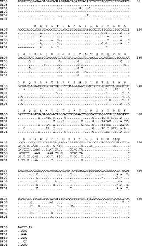
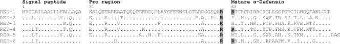
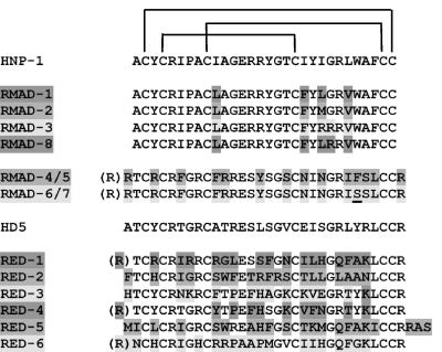
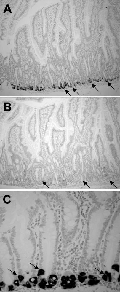
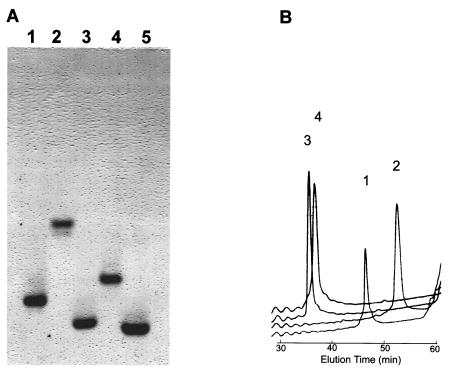
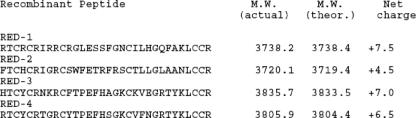
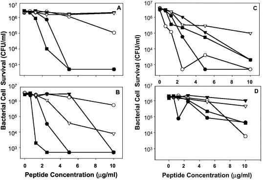
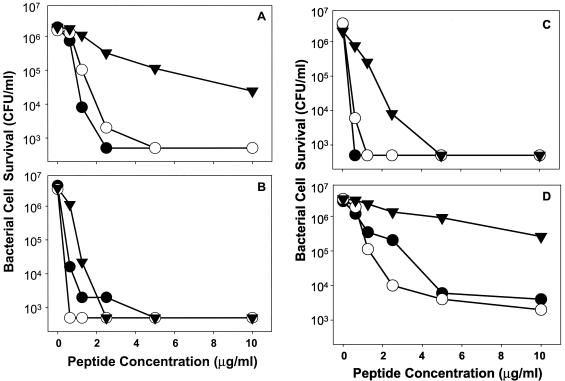
Antimicrobial peptides are secreted by small intestinal Paneth cells as components of innate immunity. To investigate the role of alpha-defensins in enteric host defenses in nonhuman primates, alpha-defensin cDNAs were isolated, alpha-defensin peptides were purified from rhesus macaque small bowel, and the bactericidal activities of the peptides were measured. Six rhesus enteric alpha-defensin (RED) cDNAs, RED-1 to RED-6, were identified in a jejunum cDNA library; the deduced RED peptides exhibited extensive diversity relative to the primary structures of rhesus myeloid alpha-defensins. RED-4 was purified from monkey jejunum, and N-terminal peptide sequencing of putative RED-4 peptides identified two N termini, RTCYCRTGR. and TCYCRTGRC.; these corresponded to alternative N termini for the RED-4 molecules, as deduced from their molecular masses and RED cDNAs. In situ hybridization experiments localized RED mRNAs exclusively to small intestinal Paneth cells. Recombinant RED-1 to RED-4 were purified to homogeneity and shown to be microbicidal in the low micromolar range (</=10 micro g/ml) against gram-positive and gram-negative bacteria, with individual peptides exhibiting variable target cell specificities. Thus, compared to myeloid alpha-defensins from rhesus macaques, enteric alpha-defensin peptides are highly variable in both primary structure and activity. These studies should facilitate further analyses of the role of alpha-defensins in primate enteric immunity.
Figures
Similar articles
-
Isolation, characterization, cDNA cloning, and antimicrobial properties of two distinct subfamilies of alpha-defensins from rhesus macaque leukocytes.Infect Immun. 1999 Nov;67(11):6139-44. doi: 10.1128/IAI.67.11.6139-6144.1999. Infect Immun. 1999. PMID: 10531277 Free PMC article.
-
alpha-Defensins from blood leukocytes of the monkey Papio hamadryas.Biochemistry (Mosc). 2006 Aug;71(8):879-83. doi: 10.1134/s0006297906080098. Biochemistry (Mosc). 2006. PMID: 16978151
-
Alpha-defensins in enteric innate immunity: functional Paneth cell alpha-defensins in mouse colonic lumen.J Biol Chem. 2009 Oct 9;284(41):27848-27856. doi: 10.1074/jbc.M109.050773. Epub 2009 Aug 17. J Biol Chem. 2009. PMID: 19687006 Free PMC article.
-
Paneth cell alpha-defensin synthesis and function.Curr Top Microbiol Immunol. 2006;306:1-25. doi: 10.1007/3-540-29916-5_1. Curr Top Microbiol Immunol. 2006. PMID: 16909916 Review.
-
Alpha-defensins in the gastrointestinal tract.Mol Immunol. 2003 Nov;40(7):463-7. doi: 10.1016/s0161-5890(03)00157-3. Mol Immunol. 2003. PMID: 14568393 Review.
Cited by
-
Strain-specific polymorphisms in Paneth cell α-defensins of C57BL/6 mice and evidence of vestigial myeloid α-defensin pseudogenes.Infect Immun. 2011 Jan;79(1):459-73. doi: 10.1128/IAI.00996-10. Epub 2010 Nov 1. Infect Immun. 2011. PMID: 21041494 Free PMC article.
-
Persistence of gut mucosal innate immune defenses by enteric α-defensin expression in the simian immunodeficiency virus model of AIDS.J Immunol. 2011 Feb 1;186(3):1589-97. doi: 10.4049/jimmunol.1002021. Epub 2010 Dec 22. J Immunol. 2011. PMID: 21178012 Free PMC article.
-
Biosynthesis and antimicrobial evaluation of backbone-cyclized α-defensins.Biochemistry. 2011 Dec 6;50(48):10508-19. doi: 10.1021/bi201430f. Epub 2011 Nov 9. Biochemistry. 2011. PMID: 22040603 Free PMC article.
-
VP4 Is a Determinant of Alpha-Defensin Modulation of Rotaviral Infection.J Virol. 2022 Apr 13;96(7):e0205321. doi: 10.1128/jvi.02053-21. Epub 2022 Mar 14. J Virol. 2022. Corrected and republished in: J Virol. 2023 Oct 31;97(10):e0096223. doi: 10.1128/jvi.00962-23. PMID: 35285683 Free PMC article. Corrected and republished.
-
Electropositive charge in alpha-defensin bactericidal activity: functional effects of Lys-for-Arg substitutions vary with the peptide primary structure.Infect Immun. 2009 Nov;77(11):5035-43. doi: 10.1128/IAI.00695-09. Epub 2009 Sep 8. Infect Immun. 2009. PMID: 19737896 Free PMC article.
References
-
- Ayabe, T., D. P. Satchell, P. Pesendorfer, H. Tanabe, C. L. Wilson, S. J. Hagen, and A. J. Ouellette. 2002. Activation of Paneth cell alpha-defensins in mouse small intestine. J. Biol. Chem. 277:5219-5228. - PubMed
-
- Ayabe, T., D. P. Satchell, C. L. Wilson, W. C. Parks, M. E. Selsted, and A. J. Ouellette. 2000. Secretion of microbicidal alpha-defensins by intestinal Paneth cells in response to bacteria. Nat. Immunol. 1:113-118. - PubMed
-
- Bevins, C. L., D. E. Jones, A. Dutra, J. Schaffzin, and M. Muenke. 1996. Human enteric defensin genes: chromosomal map position and a model for possible evolutionary relationships. Genomics 31:95-106. - PubMed
-
- Cunliffe, R. N., F. R. Rose, J. Keyte, L. Abberley, W. C. Chan, and Y. R. Mahida. 2001. Human defensin 5 is stored in precursor form in normal Paneth cells and is expressed by some villous epithelial cells and by metaplastic Paneth cells in the colon in inflammatory bowel disease. Gut 48:176-185. - PMC - PubMed
Publication types
MeSH terms
Substances
Grants and funding
LinkOut - more resources
Full Text Sources
Other Literature Sources
