. 2004 Mar;72(3):1755-9.
doi: 10.1128/IAI.72.3.1755-1759.2004.

OspB antibody prevents Borrelia burgdorferi colonization of Ixodes scapularis

Affiliations

OspB antibody prevents Borrelia burgdorferi colonization of Ixodes scapularis

Erol Fikrig et al. Infect Immun. 2004 Mar.

Abstract

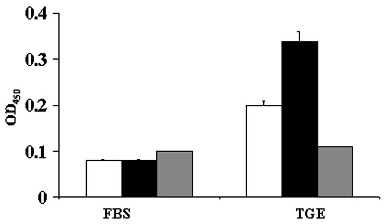
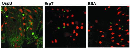
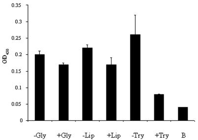
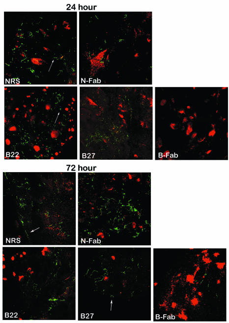
Borrelia burgdorferi outer surface protein OspB is expressed by spirochetes in the Ixodes scapularis gut. ospB is transcribed from a bicistronic operon with ospA, a known spirochete adhesion gene in the tick gut. Here we examine whether OspB also has a specific function in ticks. OspB specifically binds to a protein or protein complex within the tick gut. We also assessed whether selected nonborreliacidal OspB antibodies or F(ab)(2) fragments interfere with B. burgdorferi-tick attachment in vivo. We examined engorged ticks that fed on B. burgdorferi N40-infected scid mice that had been treated with OspB F(ab)(2) fragments. Control F(ab)(2) fragments did not interfere with B. burgdorferi colonization of the tick gut, whereas OspB F(ab)(2) fragments significantly inhibited the attachment of spirochetes to the tick gut. These studies show that nonbactericidal OspB antibodies interfere with B. burgdorferi colonization of I. scapularis, highlighting a specific role for OspB in spirochete- arthropod interactions and suggesting new antibody-mediated strategies for interfering with B. burgdorferi transmission.


Figures

FIG. 1.
FIG. 1.
OspB binds to an I. scapularis TGE. FITC-labeled OspB (black bars), ErpT (white bars) from B. burgdorferi N40, and BSA (gray bars) were used to probe either fetal bovine serum (FBS)- or TGE-coated wells. Bars represent the OD450 values at 15 min (means ± standard deviations) from three experiments. The difference between the binding of OspB to TGE and that of either BSA (P < 0.001) or ErpT (P < 0.005) was significant.
FIG. 2.
FIG. 2.
OspB directly binds to the I. scapularis gut. The direct binding of FITC-labeled OspB to the intact unfixed tick salivary gland was detected by confocal microscopy. FITC-labeled ErpT and FITC-labeled BSA were used as controls. After probing of the tick gut with FITC-labeled protein (green), the tissues were stained with propidium iodide to localize the nuclei of the gut cells (red). The FITC and propidium iodide images were examined at ×630 magnification and are presented as a single image for clarity.
FIG. 3.
FIG. 3.
OspB binding to TGE is abolished by trypsin treatment. The TGE was treated with heat-denatured glycosidases (−Gly), active glycosidases (+Gly), heat-denatured lipase (−Lip), active lipase (+Lip), soybean trypsin inhibitor and trypsin (−Try), or trypsin (+Try). The treated TGEs were then used to coat microtiter wells and probed with labeled OspB. The OD of the binding of labeled OspB to BSA is also shown as a control (B). Bars represent the OD450 values at 15 min (means ± standard deviations) from three experiments. Statistically nonsignificant differences were obtained in the cases of the glycosidase (−Gly versus +Gly)- and lipase (−Lip versus +Lip)-treated group; the difference for the protease group (−Try versus +Try) was significant (P < 0.01) by Student's t test.
FIG. 4.
FIG. 4.
OspB F(ab)2 fragments interfered with the attachment and colonization of B. burgdorferi within I. scapularis. The distribution of B. burgdorferi within the I. scapularis gut 24 and 72 h after feeding is shown. Nymphal ticks fed on B. burgdorferi-infected mice that had been treated with either normal rabbit serum (NRS), F(ab)2 fragments from normal rabbit serum (N-Fab), or OspB MAb B22 or B27 or F(ab)2 fragments prepared from polyclonal anti-OspB sera (B-Fab). The spirochetes (arrows) were stained with a FITC-labeled goat anti-B. burgdorferi antibody (green), and the nuclei of the gut epithelial cells were stained with propidium iodide (red). Images were recorded at ×400 magnification and are presented as a merged image for clarity.

Similar articles

Cited by

References

    1. Barthold, S. W., M. S. deSouza, J. L. Janotka, A. L. Smith, and D. H. Persing. 1993. Chronic Lyme borreliosis in the laboratory mouse. Am. J. Pathol. 143:959-971. - PMC - PubMed
    1. Brunet, L. R., A. Spielman, E. Fikrig, and S. R. Telford, 3rd. 1997. Heterogeneity of Lyme disease spirochaetes within individual vector ticks. Res. Microbiol. 148:437-445. - PubMed
    1. de Silva, A. M., and E. Fikrig. 1997. Arthropod- and host-specific gene expression by Borrelia burgdorferi. J. Clin. Investig. 99:377-379. - PMC - PubMed
    1. de Silva, A. M., S. R. Telford, L. R. Brunet, S. W. Barthold, and E. Fikrig. 1996. Borrelia burgdorferi OspA is an arthropod-specific transmission-blocking Lyme disease vaccine. J. Exp. Med. 183:271-275. - PMC - PubMed
    1. Escudero, R., M. L. Halluska, P. B. Backenson, J. L. Coleman, and J. L. Benach. 1997. Characterization of the physiological requirements for the bactericidal effects of a monoclonal antibody to OspB of Borrelia burgdorferi by confocal microscopy. Infect. Immun. 65:1908-1915. - PMC - PubMed

Publication types

MeSH terms

LinkOut - more resources